de Araújo Rafael B, Oyamada Maria K, Zacharias Leandro C, Cunha Leonardo P, Preti Rony C, Monteiro Mário L R
Laboratory of Investigation in Ophthalmology (LIM 33), Division of Ophthalmology, University of São Paulo Medical School, São Paulo, Brazil.
Department of Ophtalmology, School of Medicine, Federal University of Juiz de Fora, Juiz de Fora, Brazil.
Front Neurol. 2017 Dec 4;8:619. doi: 10.3389/fneur.2017.00619. eCollection 2017.
The aims of this study are to compare optical coherence tomography (OCT)-measured macular retinal layers in eyes with permanent temporal hemianopia from chiasmal compression and control eyes; to compare regular and slow-flash multifocal electroretinography (mfERG) in patients and controls; and to assess the correlation between OCT, mfERG, and central visual field (SAP) data.
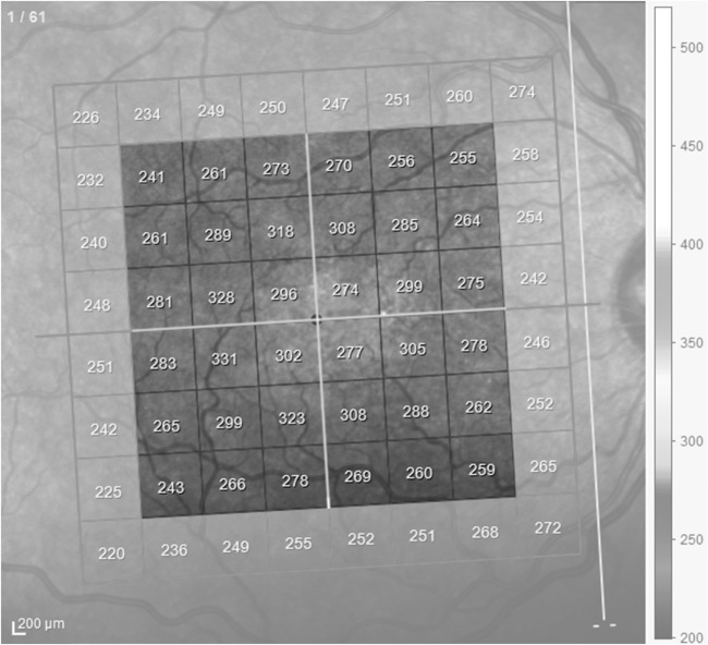
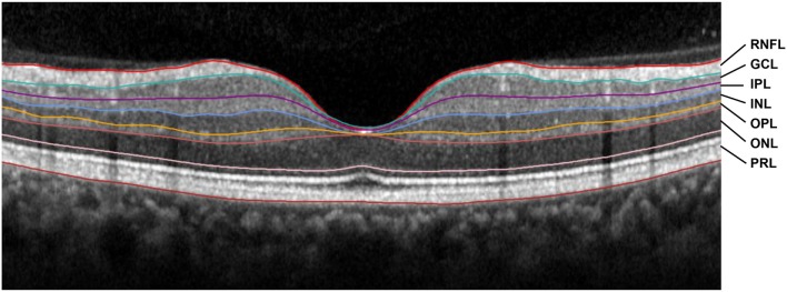
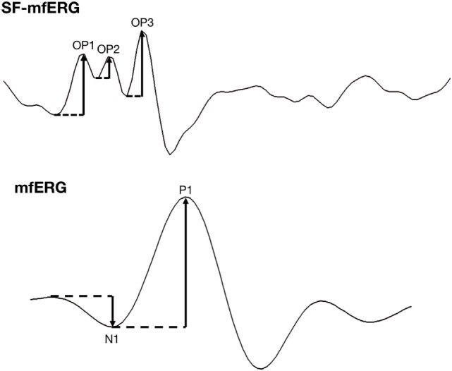
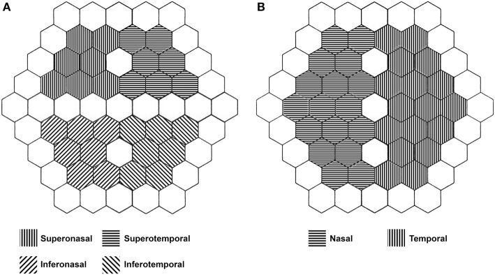
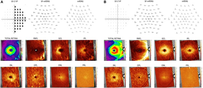
Forty-three eyes of 30 patients with permanent temporal hemianopia due to pituitary tumors who were previously submitted to chiasm decompression and 37 healthy eyes of 19 controls were submitted to macular spectral domain OCT, mfERG, and 10-2 SAP testing. After segmentation, the thickness of the macular retinal nerve fiber layer (RNFL), ganglion cell layer (GCL), inner plexiform layer (IPL), inner nuclear layer (INL), outer plexiform layer (OPL), outer nuclear layer, and photoreceptor layer (PRL) was measured. Amplitudes and oscillatory potentials (OPs) were measured on regular and slow-flash mfERG, respectively, and expressed as the mean values per quadrant and hemifield.
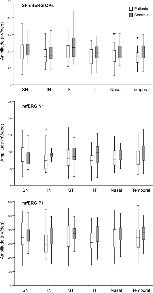
RNFL, GCL, and IPL thickness measurements were significantly reduced in all quadrants, whereas INL, OPL, and PRL thicknesses were significantly increased in the nasal quadrants in patients compared to those in controls. Significant correlations between OCT and 10-2 SAP measurements were positive for the RNFL, GCL, and IPL and negative for the INL, OPL, and PRL. OPs and mfERG N1 amplitudes were significantly reduced in the nasal hemiretina of patients. Significant correlations were found between OP and mfERG amplitudes for inner and outer nasal hemiretina OCT measurements, respectively.
Patients with permanent temporal hemianopia from previously treated chiasmal compression demonstrated significant thinning of the RNFL, GCL, IPL, and thickening of the INL, OPL, and PRL associated with reduced OP and mfERG N1 amplitudes, suggesting that axonal injury to the inner retina leads to secondary damage to the outer retina in this condition.
本研究旨在比较经光学相干断层扫描(OCT)测量的因视交叉受压导致永久性颞侧偏盲的眼睛与对照眼睛的黄斑视网膜各层;比较患者和对照者的常规和慢闪光多焦视网膜电图(mfERG);并评估OCT、mfERG和中心视野(SAP)数据之间的相关性。
对30例因垂体肿瘤导致永久性颞侧偏盲且先前接受视交叉减压术的患者的43只眼睛以及19名对照者的37只健康眼睛进行黄斑光谱域OCT、mfERG和10-2 SAP检测。分割后,测量黄斑视网膜神经纤维层(RNFL)、神经节细胞层(GCL)、内丛状层(IPL)、内核层(INL)、外丛状层(OPL)、外核层和光感受器层(PRL)的厚度。分别在常规和慢闪光mfERG上测量振幅和振荡电位(OPs),并表示为每个象限和半视野的平均值。
与对照者相比,患者所有象限的RNFL、GCL和IPL厚度测量值均显著降低,而患者鼻侧象限的INL、OPL和PRL厚度显著增加。OCT与10-2 SAP测量值之间的显著相关性在RNFL、GCL和IPL为正,在INL、OPL和PRL为负。患者鼻侧半视网膜的OPs和mfERG N1振幅显著降低。分别在鼻侧半视网膜OCT测量的内侧和外侧发现OP与mfERG振幅之间存在显著相关性。
先前接受视交叉减压术的永久性颞侧偏盲患者表现出RNFL、GCL、IPL显著变薄,INL、OPL和PRL增厚,同时OP和mfERG N1振幅降低,提示在这种情况下,视网膜内层的轴突损伤会导致外层视网膜的继发性损伤。