General Internal Medicine, Breast Diagnostic Clinic, Mayo Clinic, 200 First Street SW, Rochester, MN, USA.
Health Sciences Research, Mayo Clinic, 200 First Street SW, Rochester, MN, USA.
Breast Cancer Res. 2017 Dec 19;19(1):134. doi: 10.1186/s13058-017-0922-6.
Over 40% of women undergoing breast screening have mammographically dense breasts. Elevated mammographic breast density (MBD) is an established breast cancer risk factor and is known to mask tumors within the dense tissue. However, the association of MBD with high risk benign breast disease (BBD) is unknown.
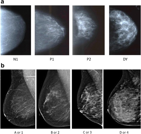
We analyzed data for 3400 women diagnosed with pathologically confirmed BBD in the Mayo Clinic BBD cohort from 1985-2001, with a clinical MBD measure (either parenchymal pattern (PP) or Breast Imaging Reporting and Data Systems (BI-RADS) density) and expert pathology review. Risk factor information was collected from medical records and questionnaires. MBD was dichotomized as dense (PP classification P2 or DY, or BI-RADS classification c or d) or non-dense (PP classification N1 or P1, or BI-RADS classification a or b). Associations of clinical and histologic characteristics with MBD were examined using logistic regression analysis to estimate odds ratios (ORs) with 95% confidence intervals (CIs).
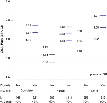
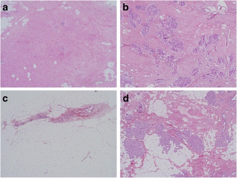
Of 3400 women in the study, 2163 (64%) had dense breasts. Adjusting for age and body mass index (BMI), there were positive associations of dense breasts with use of hormone therapy (HT), lack of lobular involution, presence of atypical lobular hyperplasia (ALH), histologic fibrosis, columnar cell hyperplasia/flat epithelia atypia (CCH/FEA), sclerosing adenosis (SA), cyst, usual ductal hyperplasia, and calcifications. In fully adjusted multivariate models, HT (1.3, 95% CI 1.1-1.5), ALH (1.5, 95% CI 1.0-2.2), lack of lobular involution (OR 1.6, 95% CI 1.2-2.1, compared to complete involution), fibrosis (OR 2.2, 95% CI 1.9-2.6) and CCH/FEA (OR 1.3, 95% CI 1.0-1.6) remained significantly associated with high MBD.
Our findings support an association between high risk BBD and high MBD, suggesting that risks associated with the latter may act early in breast carcinogenesis.
超过 40%接受乳房筛查的女性乳房 X 光片显示致密。乳腺 X 光片致密(MBD)是乳腺癌的一个既定危险因素,并且已知会掩盖致密组织内的肿瘤。然而,MBD 与高风险良性乳腺疾病(BBD)的关联尚不清楚。
我们分析了 1985 年至 2001 年在 Mayo 诊所 BBD 队列中诊断为病理证实的 BBD 的 3400 名女性的数据,这些女性具有临床 MBD 测量值(实质模式(PP)或乳腺成像报告和数据系统(BI-RADS)密度)和专家病理审查。危险因素信息从病历和问卷中收集。MBD 分为致密(PP 分类 P2 或 DY,或 BI-RADS 分类 c 或 d)或非致密(PP 分类 N1 或 P1,或 BI-RADS 分类 a 或 b)。使用逻辑回归分析检查临床和组织学特征与 MBD 的关联,以估计比值比(OR)及其 95%置信区间(CI)。
在研究的 3400 名女性中,2163 名(64%)乳房致密。调整年龄和体重指数(BMI)后,致密乳房与激素治疗(HT)的使用、小叶不完整、存在非典型小叶增生(ALH)、组织纤维化、柱状细胞增生/扁平上皮不典型(CCH/FEA)、硬化性腺病(SA)、囊肿、普通导管增生和钙化呈正相关。在完全调整的多变量模型中,HT(1.3,95%CI 1.1-1.5)、ALH(1.5,95%CI 1.0-2.2)、小叶不完整(与完全不完整相比,OR 1.6,95%CI 1.2-2.1)、纤维化(OR 2.2,95%CI 1.9-2.6)和 CCH/FEA(OR 1.3,95%CI 1.0-1.6)与高 MBD 仍显著相关。
我们的研究结果支持高风险 BBD 与高 MBD 之间的关联,表明后者相关的风险可能在乳腺癌发生的早期发挥作用。