Center for Transdisciplinary Research, Institute for Research Promotion, Niigata University, Niigata, 951-8510, Japan.
Department of Neurochemistry and Molecular Cell Biology, Graduate School of Medical and Dental Sciences, Niigata University, Niigata, 951-8510, Japan.
Nat Commun. 2017 Dec 19;8(1):2194. doi: 10.1038/s41467-017-02193-w.
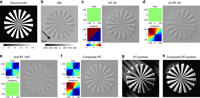
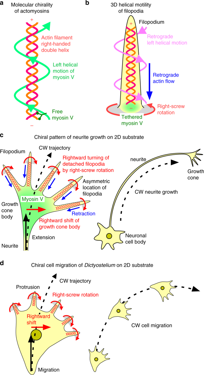
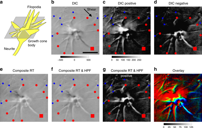
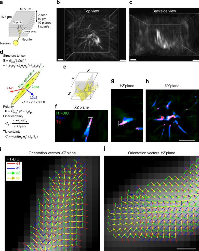
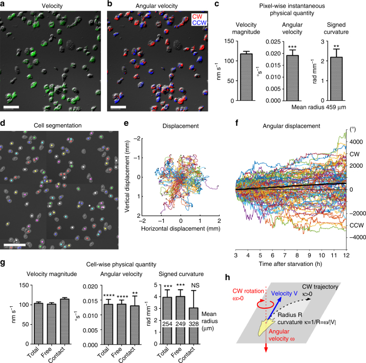
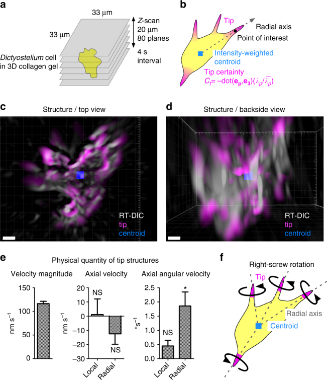
Left-right asymmetry is a fundamental feature of body plans, but its formation mechanisms and roles in functional lateralization remain unclear. Accumulating evidence suggests that left-right asymmetry originates in the cellular chirality. However, cell chirality has not yet been quantitatively investigated, mainly due to the absence of appropriate methods. Here we combine 3D Riesz transform-differential interference contrast (RT-DIC) microscopy and computational kinematic analysis to characterize chiral cellular morphology and motility. We reveal that filopodia of neuronal growth cones exhibit 3D left-helical motion with retraction and right-screw rotation. We next apply the methods to amoeba Dictyostelium discoideum and discover right-handed clockwise cell migration on a 2D substrate and right-screw rotation of subcellular protrusions along the radial axis in a 3D substrate. Thus, RT-DIC microscopy and the computational kinematic analysis are useful and versatile tools to reveal the mechanisms of left-right asymmetry formation and the emergence of lateralized functions.
左右不对称是身体结构的一个基本特征,但它的形成机制及其在功能偏侧化中的作用仍不清楚。越来越多的证据表明,左右不对称起源于细胞的手性。然而,由于缺乏适当的方法,细胞手性尚未得到定量研究。在这里,我们结合 3D Riesz 变换-微分干涉对比(RT-DIC)显微镜和计算运动学分析来描述手性细胞形态和运动。我们揭示神经元生长锥的丝状伪足表现出具有回缩和右旋螺旋旋转的 3D 左旋运动。接下来,我们将该方法应用于变形虫 Dictyostelium discoideum,并发现细胞在二维基质上的右旋顺时针迁移以及在三维基质中沿着放射轴的亚细胞突起的右旋螺旋旋转。因此,RT-DIC 显微镜和计算运动学分析是揭示左右不对称形成机制和出现偏侧化功能的有用和通用工具。