Mauland Karen Klepsland, Eng Øyvin, Ytre-Hauge Sigmund, Tangen Ingvild L, Berg Anna, Salvesen Helga B, Salvesen Øyvind O, Krakstad Camilla, Trovik Jone, Hoivik Erling A, Werner Henrica Maria Johanna, Mellgren Gunnar, Haldorsen Ingfrid S
Centre for Cancer Biomarkers, CCBIO, Department of Clinical Science (K2), University of Bergen, Bergen, Norway.
Department of Gynecology and Obstetrics, Haukeland University Hospital, Bergen, Norway.
Oncotarget. 2017 Oct 19;8(62):105184-105195. doi: 10.18632/oncotarget.21917. eCollection 2017 Dec 1.
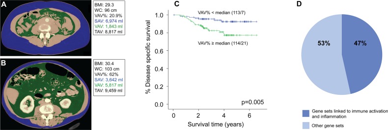
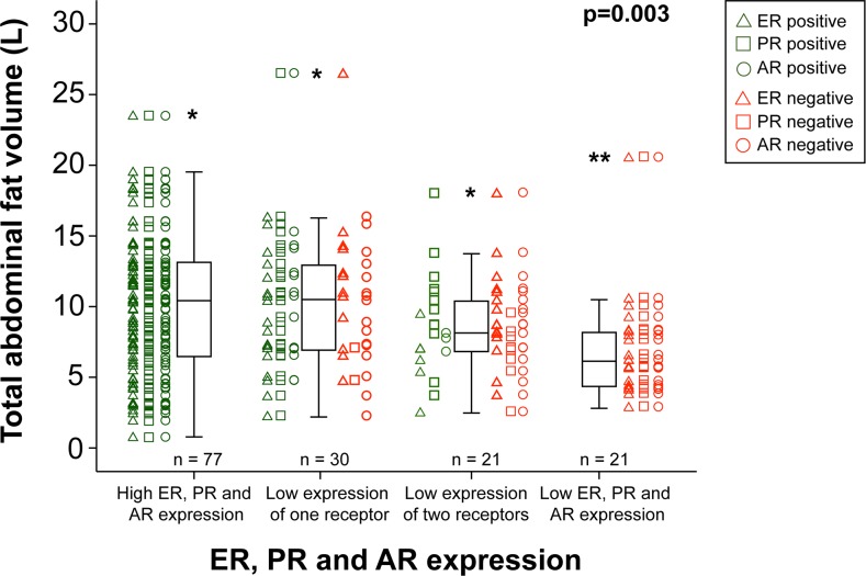
Despite evidence of increased endometrial cancer (EC) risk in obese women, the impact of obesity on clinical and histological phenotype is poorly understood. This study explored abdominal fat volumes and fat distribution quantified by computed tomography (CT), in relation to tumor characteristics and outcome. 227 EC patients with preoperative abdominal CT scans were included. Total abdominal fat volume (TAV), subcutaneous abdominal fat volume (SAV) and visceral abdominal fat volume (VAV) were quantified, and visceral fat percentage calculated (VAV%=[VAV/TAV]x100). Waist circumference (WC) and liver density (LD) were measured, and body mass index (BMI) calculated. Data for estrogen, progesterone and androgen receptor (ERα/PR/AR) expression by immunohistochemistry were available for 149 tumors, and global gene expression data for 105 tumors. High BMI, TAV, SAV, VAV and WC, and low LD, were associated with low grade endometrioid tumors and PR and AR positivity (all p≤0.03). High VAV% was associated with high age (p<0.001), aneuploidy (p=0.01) and independently predicted reduced disease-specific survival (HR 1.05, 95% CI 1.00-1.11, p=0.041). Tumors from patients with low VAV% showed enrichment of gene sets related to immune activation and inflammation. In conclusion, high VAV% independently predicts reduced EC survival. Tumors arising in patients with low VAV% show enrichment of immune and inflammation related gene sets, suggesting that the global metabolic setting may be important for tumor immune response.
尽管有证据表明肥胖女性患子宫内膜癌(EC)的风险增加,但肥胖对临床和组织学表型的影响却知之甚少。本研究探讨了通过计算机断层扫描(CT)量化的腹部脂肪体积和脂肪分布与肿瘤特征及预后的关系。纳入了227例术前行腹部CT扫描的EC患者。对腹部总脂肪体积(TAV)、皮下腹部脂肪体积(SAV)和内脏腹部脂肪体积(VAV)进行量化,并计算内脏脂肪百分比(VAV%=[VAV/TAV]×100)。测量腰围(WC)和肝脏密度(LD),并计算体重指数(BMI)。149个肿瘤可获得免疫组化检测雌激素、孕激素和雄激素受体(ERα/PR/AR)表达的数据,105个肿瘤可获得全基因表达数据。高BMI、TAV、SAV、VAV和WC以及低LD与低级别子宫内膜样肿瘤以及PR和AR阳性相关(所有p≤0.03)。高VAV%与高龄(p<0.001)、非整倍体(p=0.01)相关,并且独立预测疾病特异性生存率降低(HR 1.05,95%CI 1.00-1.11,p=0.041)。VAV%低的患者的肿瘤显示与免疫激活和炎症相关的基因集富集。总之,高VAV%独立预测EC生存率降低。VAV%低的患者发生的肿瘤显示免疫和炎症相关基因集富集,这表明整体代谢环境可能对肿瘤免疫反应很重要。