Terunuma Toshiyuki, Tokui Aoi, Sakae Takeji
Faculty of Medicine, University of Tsukuba, Ten-nohdai 1-1-1, Tsukuba, 305-8575, Japan.
Proton Medical Research Center (PMRC), University of Tsukuba Hospital, Amakubo 2-1-1, Tsukuba, 305-8576, Japan.
Radiol Phys Technol. 2018 Mar;11(1):43-53. doi: 10.1007/s12194-017-0435-0. Epub 2017 Dec 28.
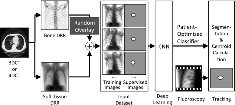
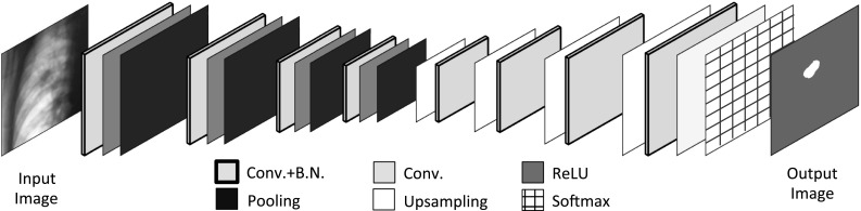
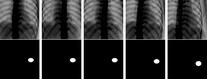
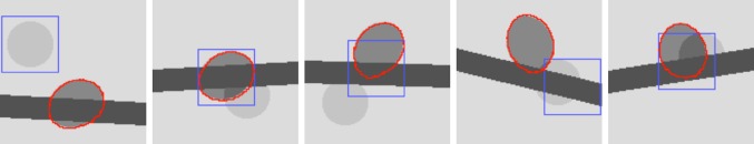
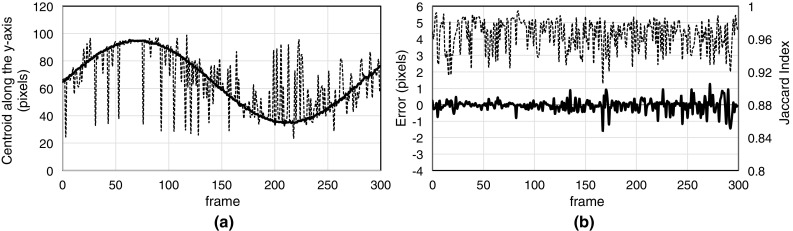
Robustness to obstacles is the most important factor necessary to achieve accurate tumor tracking without fiducial markers. Some high-density structures, such as bone, are enhanced on X-ray fluoroscopic images, which cause tumor mistracking. Tumor tracking should be performed by controlling "importance recognition": the understanding that soft-tissue is an important tracking feature and bone structure is unimportant. We propose a new real-time tumor-contouring method that uses deep learning with importance recognition control. The novelty of the proposed method is the combination of the devised random overlay method and supervised deep learning to induce the recognition of structures in tumor contouring as important or unimportant. This method can be used for tumor contouring because it uses deep learning to perform image segmentation. Our results from a simulated fluoroscopy model showed accurate tracking of a low-visibility tumor with an error of approximately 1 mm, even if enhanced bone structure acted as an obstacle. A high similarity of approximately 0.95 on the Jaccard index was observed between the segmented and ground truth tumor regions. A short processing time of 25 ms was achieved. The results of this simulated fluoroscopy model support the feasibility of robust real-time tumor contouring with fluoroscopy. Further studies using clinical fluoroscopy are highly anticipated.
对障碍物的鲁棒性是在没有基准标记的情况下实现精确肿瘤跟踪所必需的最重要因素。一些高密度结构,如骨骼,在X射线荧光透视图像上会增强,这会导致肿瘤跟踪错误。肿瘤跟踪应通过控制“重要性识别”来进行:即认识到软组织是重要的跟踪特征,而骨骼结构不重要。我们提出了一种新的实时肿瘤轮廓绘制方法,该方法使用带有重要性识别控制的深度学习。所提出方法的新颖之处在于将设计的随机叠加方法与监督深度学习相结合,以在肿瘤轮廓绘制中引导对结构重要性或不重要性的识别。该方法可用于肿瘤轮廓绘制,因为它使用深度学习来执行图像分割。我们在模拟荧光透视模型中的结果表明,即使增强的骨骼结构成为障碍物,也能以约1毫米的误差精确跟踪低可见度肿瘤。在分割的肿瘤区域和真实肿瘤区域之间观察到杰卡德指数约为0.95的高度相似性。实现了25毫秒的短处理时间。该模拟荧光透视模型的结果支持了使用荧光透视进行鲁棒实时肿瘤轮廓绘制的可行性。非常期待使用临床荧光透视的进一步研究。