Department of Cardiology, Renmin Hospital of Wuhan University, Cardiovascular Research Institute, Wuhan University, Hubei Key Laboratory of Cardiology, Wuhan, Hubei 430060, P.R. China.
Int J Mol Med. 2018 Mar;41(3):1265-1274. doi: 10.3892/ijmm.2017.3351. Epub 2017 Dec 29.
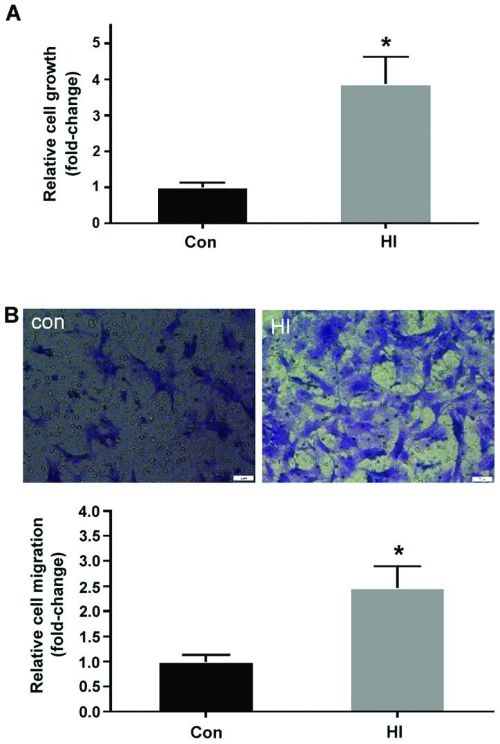
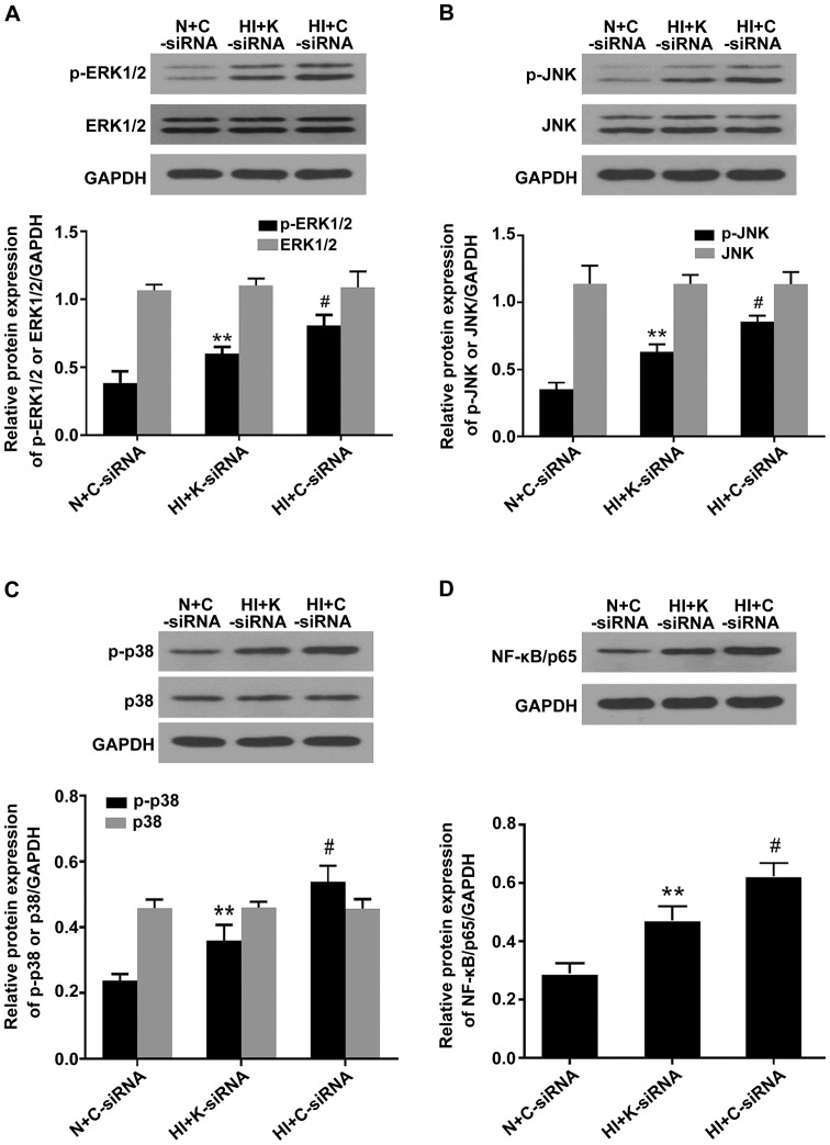
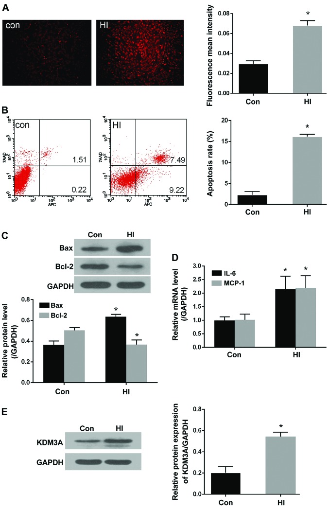
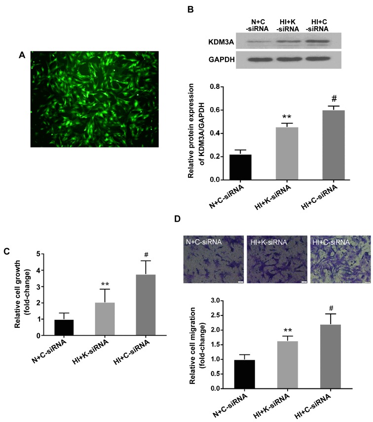
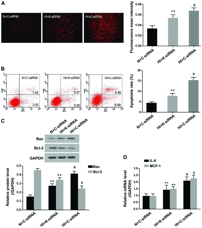
Previous studies have indicated that lysine (K)‑specific demethylase 3A (KDM3A) is associated with diverse diabetes‑associated cardiovascular complications in response to high glucose levels. However, the effects of KDM3A on the pathological progression of cardiovascular injuries in response to high insulin levels remain unknown. The present study aimed to explore whether KDM3A knockdown may attenuate high insulin‑induced vascular smooth muscle cell (VSMC) dysfunction, and to further investigate the underlying mechanisms. Primary VSMCs were isolated from the thoracic aorta of Sprague‑Dawley rats. Lentiviral vectors encoding control‑small interfering (si)RNA or KDM3A‑siRNA were transduced into VSMCs for 72 h, and cells were subsequently incubated in medium containing 100 nM insulin for a further 5 days. Cellular proli-feration, migration and apoptosis were measured by Cell Counting kit‑8, Transwell chamber assay and flow cytometry, respectively. Reactive oxygen species (ROS) were detected using the dihydroethidium fluorescent probe. The mRNA expression levels of interleukin‑6 and monocyte chemotactic protein‑1 were measured by reverse transcription‑quantitative polymerase chain reaction. Furthermore, the protein expression levels of KDM3A, mitogen‑activated protein kinases (MAPKs), nuclear factor (NF)‑κB/p65, B‑cell lymphoma 2 (Bcl‑2)‑associated X protein and Bcl‑2 were evaluated by west-ern blotting. Lentivirus transduction with KDM3A‑siRNA markedly reduced the elevated expression of KDM3A induced by high insulin stimulation in VSMCs. In addition, inhibition of KDM3A significantly ameliorated insulin‑induced VSMC proliferation and migration, which was accompanied by decreased ROS levels, cell apoptosis and inflammatory cytokine levels. Furthermore, KDM3A gene silencing mitigated phosphorylation of MAPKs and NF‑κB/p65 activation. In conclusion, KDM3A inhibition may exert numerous protective effects on high insulin‑stimulated VSMCs, and the underlying mechanisms may be partly associated with inactivation of MAPK/NF‑κB signaling pathways.
先前的研究表明,赖氨酸特异性脱甲基酶 3A(KDM3A)与高糖水平下多种与糖尿病相关的心血管并发症有关。然而,KDM3A 对高胰岛素水平引起的心血管损伤病理进展的影响尚不清楚。本研究旨在探讨 KDM3A 敲低是否可以减轻高胰岛素诱导的血管平滑肌细胞(VSMC)功能障碍,并进一步探讨其潜在机制。从 Sprague-Dawley 大鼠的胸主动脉中分离原代 VSMC。将编码对照小干扰(si)RNA 或 KDM3A-siRNA 的慢病毒载体转染至 VSMC 中 72 h,随后将细胞在含有 100 nM 胰岛素的培养基中孵育 5 天。通过细胞计数试剂盒-8、Transwell 室测定和流式细胞术分别测量细胞增殖、迁移和凋亡。使用二氢乙啶荧光探针检测活性氧(ROS)。通过逆转录-定量聚合酶链反应测量白细胞介素-6 和单核细胞趋化蛋白-1 的 mRNA 表达水平。此外,通过 Western blot 评估 KDM3A、丝裂原活化蛋白激酶(MAPKs)、核因子(NF)-κB/p65、B 细胞淋巴瘤 2(Bcl-2)相关 X 蛋白和 Bcl-2 的蛋白表达水平。KDM3A-siRNA 的慢病毒转导显著降低了高胰岛素刺激诱导的 VSMC 中 KDM3A 的上调表达。此外,抑制 KDM3A 显著改善了胰岛素诱导的 VSMC 增殖和迁移,同时降低了 ROS 水平、细胞凋亡和炎症细胞因子水平。此外,KDM3A 基因沉默减轻了 MAPK/NF-κB 信号通路的激活。综上所述,KDM3A 抑制可能对高胰岛素刺激的 VSMC 发挥多种保护作用,其潜在机制可能部分与 MAPK/NF-κB 信号通路失活有关。