Lin Cui, Zhu Dongxing, Markby Greg, Corcoran Brendan M, Farquharson Colin, Macrae Vicky E
Developmental Biology, The Roslin Institute and R(D)SVS, University of Edinburgh;
Guangzhou Institute of Cardiovascular Disease, The Second Affiliated Hospital, School of Basic Medical Sciences, Guangzhou Medical University.
J Vis Exp. 2017 Nov 20(129):56126. doi: 10.3791/56126.
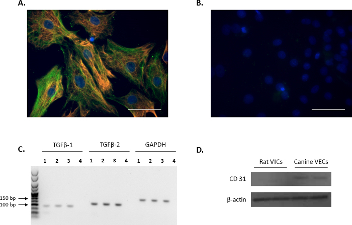
Calcific aortic valve disease (CAVD) is characterized by the progressive thickening of the aortic valve leaflets. It is a condition frequently found in the elderly and end-stage renal disease (ESRD) patients, who commonly suffer from hyperphosphatemia and hypercalcemia. At present, there are no medication therapies that can stop its progression. The mechanisms that underlie this pathological process remain unclear. The aortic valve leaflet is composed of a thin layer of valve endothelial cells (VECs) on the outer surfaces of the aortic cusps, with valve interstitial cells (VICs) sandwiched between the VECs. The use of a rat model enables the in vitro study of ectopic calcification based on the in vivo physiopathological serum phosphate (Pi) and calcium (Ca) levels of patients who suffer from hyperphosphatemia and hypercalcemia. The described protocol details the isolation of a pure rat VIC population as shown by the expression of VIC markers: alpha-smooth muscle actin (α-SMA) vimentin and tissue growth factor beta (TGFβ) 1 and 2, and the absence of cluster of differentiation (CD) 31, a VEC marker. By expanding these VICs, biochemical, genetic, and imaging studies can be performed to study and unravel the key mediators underpinning CAVD.
钙化性主动脉瓣疾病(CAVD)的特征是主动脉瓣小叶逐渐增厚。它是一种在老年人和终末期肾病(ESRD)患者中常见的病症,这些患者通常患有高磷血症和高钙血症。目前,尚无能够阻止其进展的药物疗法。这种病理过程的潜在机制仍不清楚。主动脉瓣小叶由主动脉瓣叶外表面的一层薄的瓣膜内皮细胞(VECs)组成,瓣膜间质细胞(VICs)夹在VECs之间。使用大鼠模型能够基于患有高磷血症和高钙血症患者的体内生理病理血清磷酸盐(Pi)和钙(Ca)水平对异位钙化进行体外研究。所描述的方案详细说明了纯大鼠VIC群体的分离,如通过VIC标志物的表达所示:α-平滑肌肌动蛋白(α-SMA)、波形蛋白和组织生长因子β(TGFβ)1和2,以及不存在作为VEC标志物的分化簇(CD)31。通过扩增这些VICs,可以进行生化、遗传和成像研究,以研究和揭示CAVD的关键介质。