• 文献检索
  • 文档翻译
  • 深度研究
  • 学术资讯
  • Suppr Zotero 插件Zotero 插件
  • 邀请有礼
  • 套餐&价格
  • 历史记录
应用&插件
Suppr Zotero 插件Zotero 插件浏览器插件Mac 客户端Windows 客户端微信小程序
定价
高级版会员购买积分包购买API积分包
服务
文献检索文档翻译深度研究API 文档MCP 服务
关于我们
关于 Suppr公司介绍联系我们用户协议隐私条款
关注我们

Suppr 超能文献

核心技术专利:CN118964589B侵权必究
粤ICP备2023148730 号-1Suppr @ 2026

文献检索

告别复杂PubMed语法,用中文像聊天一样搜索,搜遍4000万医学文献。AI智能推荐,让科研检索更轻松。

立即免费搜索

文件翻译

保留排版,准确专业,支持PDF/Word/PPT等文件格式,支持 12+语言互译。

免费翻译文档

深度研究

AI帮你快速写综述,25分钟生成高质量综述,智能提取关键信息,辅助科研写作。

立即免费体验

合成心脏瓣膜组织工程支架的开发策略

Strategies for Development of Synthetic Heart Valve Tissue Engineering Scaffolds.

作者信息

Snyder Yuriy, Jana Soumen

机构信息

Department of Bioengineering, University of Missouri, Columbia, MO 65211, USA.

出版信息

Prog Mater Sci. 2023 Oct;139. doi: 10.1016/j.pmatsci.2023.101173. Epub 2023 Jul 26.

DOI:10.1016/j.pmatsci.2023.101173
PMID:37981978
原文链接:https://pmc.ncbi.nlm.nih.gov/articles/PMC10655624/
Abstract

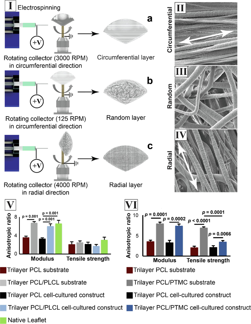
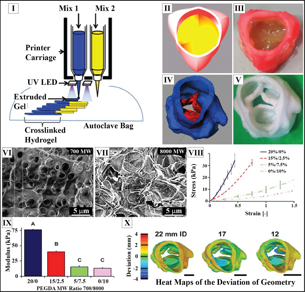
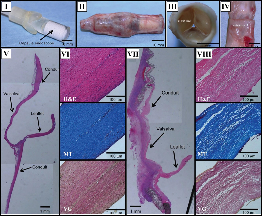
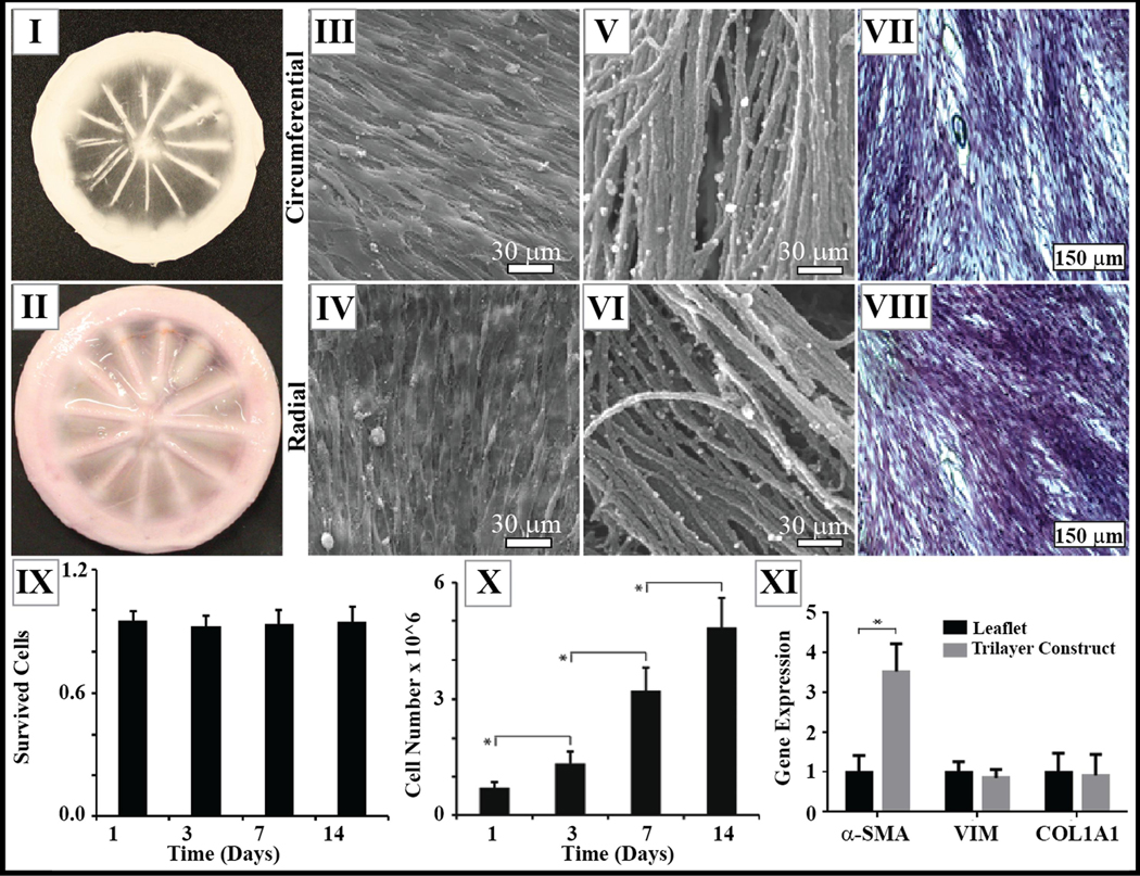
The current clinical solutions, including mechanical and bioprosthetic valves for valvular heart diseases, are plagued by coagulation, calcification, nondurability, and the inability to grow with patients. The tissue engineering approach attempts to resolve these shortcomings by producing heart valve scaffolds that may deliver patients a life-long solution. Heart valve scaffolds serve as a three-dimensional support structure made of biocompatible materials that provide adequate porosity for cell infiltration, and nutrient and waste transport, sponsor cell adhesion, proliferation, and differentiation, and allow for extracellular matrix production that together contributes to the generation of functional neotissue. The foundation of successful heart valve tissue engineering is replicating native heart valve architecture, mechanics, and cellular attributes through appropriate biomaterials and scaffold designs. This article reviews biomaterials, the fabrication of heart valve scaffolds, and their in-vitro and in-vivo evaluations applied for heart valve tissue engineering.

摘要

目前的临床解决方案,包括用于心脏瓣膜疾病的机械瓣膜和生物人工瓣膜,都存在凝血、钙化、耐久性差以及无法随患者生长等问题。组织工程方法试图通过制造心脏瓣膜支架来解决这些缺点,这些支架可能为患者提供终身解决方案。心脏瓣膜支架是由生物相容性材料制成的三维支撑结构,它为细胞浸润、营养物质和废物运输提供足够的孔隙率,促进细胞黏附、增殖和分化,并允许细胞外基质产生,共同促进功能性新组织的生成。成功的心脏瓣膜组织工程的基础是通过适当的生物材料和支架设计来复制天然心脏瓣膜的结构、力学和细胞特性。本文综述了用于心脏瓣膜组织工程的生物材料、心脏瓣膜支架的制造及其体外和体内评估。

https://cdn.ncbi.nlm.nih.gov/pmc/blobs/6f96/10655624/4335f9e3af42/nihms-1922958-f0024.jpg
https://cdn.ncbi.nlm.nih.gov/pmc/blobs/6f96/10655624/c104c2a07f65/nihms-1922958-f0001.jpg
https://cdn.ncbi.nlm.nih.gov/pmc/blobs/6f96/10655624/482fe2be7587/nihms-1922958-f0002.jpg
https://cdn.ncbi.nlm.nih.gov/pmc/blobs/6f96/10655624/5eb8d2c3b6aa/nihms-1922958-f0003.jpg
https://cdn.ncbi.nlm.nih.gov/pmc/blobs/6f96/10655624/d4f3738b959f/nihms-1922958-f0004.jpg
https://cdn.ncbi.nlm.nih.gov/pmc/blobs/6f96/10655624/5224dde7d1c2/nihms-1922958-f0005.jpg
https://cdn.ncbi.nlm.nih.gov/pmc/blobs/6f96/10655624/2942bda2164e/nihms-1922958-f0006.jpg
https://cdn.ncbi.nlm.nih.gov/pmc/blobs/6f96/10655624/8d0cd5dcdf46/nihms-1922958-f0007.jpg
https://cdn.ncbi.nlm.nih.gov/pmc/blobs/6f96/10655624/776f3e7dc6c2/nihms-1922958-f0008.jpg
https://cdn.ncbi.nlm.nih.gov/pmc/blobs/6f96/10655624/df8f9d79cedd/nihms-1922958-f0009.jpg
https://cdn.ncbi.nlm.nih.gov/pmc/blobs/6f96/10655624/6740c8030b44/nihms-1922958-f0010.jpg
https://cdn.ncbi.nlm.nih.gov/pmc/blobs/6f96/10655624/2bbf39c2c198/nihms-1922958-f0011.jpg
https://cdn.ncbi.nlm.nih.gov/pmc/blobs/6f96/10655624/7f31b6d4baf2/nihms-1922958-f0012.jpg
https://cdn.ncbi.nlm.nih.gov/pmc/blobs/6f96/10655624/ae8214f2f120/nihms-1922958-f0013.jpg
https://cdn.ncbi.nlm.nih.gov/pmc/blobs/6f96/10655624/decaf01d0b95/nihms-1922958-f0014.jpg
https://cdn.ncbi.nlm.nih.gov/pmc/blobs/6f96/10655624/d50011dab776/nihms-1922958-f0015.jpg
https://cdn.ncbi.nlm.nih.gov/pmc/blobs/6f96/10655624/5c83612668a8/nihms-1922958-f0016.jpg
https://cdn.ncbi.nlm.nih.gov/pmc/blobs/6f96/10655624/29f8a17d8c64/nihms-1922958-f0017.jpg
https://cdn.ncbi.nlm.nih.gov/pmc/blobs/6f96/10655624/0f7756043454/nihms-1922958-f0018.jpg
https://cdn.ncbi.nlm.nih.gov/pmc/blobs/6f96/10655624/70d52bbdc36a/nihms-1922958-f0019.jpg
https://cdn.ncbi.nlm.nih.gov/pmc/blobs/6f96/10655624/f25eb78a8163/nihms-1922958-f0020.jpg
https://cdn.ncbi.nlm.nih.gov/pmc/blobs/6f96/10655624/2ae8d2560298/nihms-1922958-f0021.jpg
https://cdn.ncbi.nlm.nih.gov/pmc/blobs/6f96/10655624/7f0830a23a23/nihms-1922958-f0022.jpg
https://cdn.ncbi.nlm.nih.gov/pmc/blobs/6f96/10655624/001a6b58fd8b/nihms-1922958-f0023.jpg
https://cdn.ncbi.nlm.nih.gov/pmc/blobs/6f96/10655624/4335f9e3af42/nihms-1922958-f0024.jpg

相似文献

1
Strategies for Development of Synthetic Heart Valve Tissue Engineering Scaffolds.合成心脏瓣膜组织工程支架的开发策略
Prog Mater Sci. 2023 Oct;139. doi: 10.1016/j.pmatsci.2023.101173. Epub 2023 Jul 26.
2
Modifying decellularized aortic valve scaffolds with stromal cell-derived factor-1α loaded proteolytically degradable hydrogel for recellularization and remodeling.用负载基质细胞衍生因子-1α的蛋白水解可降解水凝胶修饰去细胞主动脉瓣支架以进行再细胞化和重塑。
Acta Biomater. 2019 Apr 1;88:280-292. doi: 10.1016/j.actbio.2019.02.002. Epub 2019 Feb 2.
3
Strategies for development of decellularized heart valve scaffolds for tissue engineering.用于组织工程的脱细胞心脏瓣膜支架的开发策略。
Biomaterials. 2022 Sep;288:121675. doi: 10.1016/j.biomaterials.2022.121675. Epub 2022 Jul 18.
4
Scaffolds for tissue engineering of cardiac valves.用于心脏瓣膜组织工程的支架。
Acta Biomater. 2014 Jul;10(7):2877-93. doi: 10.1016/j.actbio.2014.03.014. Epub 2014 Mar 24.
5
Living nano-micro fibrous woven fabric/hydrogel composite scaffolds for heart valve engineering.用于心脏瓣膜工程的活性纳米-微纤维编织织物/水凝胶复合支架
Acta Biomater. 2017 Mar 15;51:89-100. doi: 10.1016/j.actbio.2017.01.051. Epub 2017 Jan 18.
6
Fiber-reinforced hydrogel scaffolds for heart valve tissue engineering.用于心脏瓣膜组织工程的纤维增强水凝胶支架
J Biomater Appl. 2014 Sep;29(3):399-410. doi: 10.1177/0885328214530589. Epub 2014 Apr 14.
7
Engineering of a polymer layered bio-hybrid heart valve scaffold.聚合物层状生物杂交心脏瓣膜支架的工程设计。
Mater Sci Eng C Mater Biol Appl. 2015 Jun;51:263-73. doi: 10.1016/j.msec.2015.03.009. Epub 2015 Mar 11.
8
Melt-electrowriting-enabled anisotropic scaffolds loaded with valve interstitial cells for heart valve tissue Engineering.熔融静电纺丝法制备负载心脏瓣膜间质细胞的各向异性支架用于心脏瓣膜组织工程。
J Nanobiotechnology. 2024 Jun 28;22(1):378. doi: 10.1186/s12951-024-02656-5.
9
Decellularized tissue-engineered heart valves calcification: what do animal and clinical studies tell us?去细胞组织工程心脏瓣膜钙化:动物和临床研究告诉了我们什么?
J Mater Sci Mater Med. 2020 Dec 5;31(12):132. doi: 10.1007/s10856-020-06462-x.
10
Leaflet Tissue Generation from Microfibrous Heart Valve Leaflet Scaffolds with Native Characteristics.具有天然特性的微纤维心脏瓣膜叶瓣支架的小叶片组织生成。
ACS Appl Bio Mater. 2021 Nov 15;4(11):7836-7847. doi: 10.1021/acsabm.1c00768. Epub 2021 Nov 3.

引用本文的文献

1
Biodegradable Polyesters: Approaches to Increase Degradation Rates for Biomedical Applications.可生物降解聚酯:提高生物医学应用降解速率的方法
ACS Macro Lett. 2025 Aug 19;14(8):1221-1240. doi: 10.1021/acsmacrolett.5c00417. Epub 2025 Aug 10.
2
Mechanistic Insights into Bioprosthetic Heart Valve Calcification and Anti-Calcification Strategies.生物人工心脏瓣膜钙化及抗钙化策略的机制性见解
Rev Cardiovasc Med. 2025 May 20;26(5):36688. doi: 10.31083/RCM36688. eCollection 2025 May.
3
A chronological history of heart valve prostheses to offer perspectives of their limitations.

本文引用的文献

1
Calcification in Pulmonary Heart Valve Tissue Engineering: A Systematic Review and Meta-Analysis of Large-Animal Studies.肺动脉瓣组织工程中的钙化:大型动物研究的系统评价与荟萃分析
JACC Basic Transl Sci. 2023 Jan 25;8(5):572-591. doi: 10.1016/j.jacbts.2022.09.009. eCollection 2023 May.
2
Designing an Innovative Electrospinning Strategy to Generate PHBV Nanofiber Scaffolds with a Radially Oriented Fibrous Pattern.设计一种创新的静电纺丝策略以制备具有径向取向纤维图案的聚(3-羟基丁酸酯-co-3-羟基戊酸酯)纳米纤维支架。
Nanomaterials (Basel). 2023 Mar 23;13(7):1150. doi: 10.3390/nano13071150.
3
Polymeric prosthetic heart valves: A review of current technologies and future directions.
心脏瓣膜假体的编年历史,以展现其局限性。
Front Bioeng Biotechnol. 2025 Feb 14;13:1533421. doi: 10.3389/fbioe.2025.1533421. eCollection 2025.
4
A self-sacrificing anti-inflammatory coating promotes simultaneous cardiovascular repair and reendothelialization of implanted devices.一种自我牺牲的抗炎涂层可促进植入装置的心血管修复和再内皮化同步进行。
Bioact Mater. 2025 Feb 13;47:502-512. doi: 10.1016/j.bioactmat.2025.01.037. eCollection 2025 May.
5
Innovative hydrogel solutions for articular cartilage regeneration: a comprehensive review.用于关节软骨再生的创新水凝胶解决方案:全面综述。
Int J Surg. 2024 Dec 1;110(12):7984-8001. doi: 10.1097/JS9.0000000000002076.
6
Non-immune factors cause prolonged myofibroblast phenotype in implanted synthetic heart valve scaffolds.非免疫因素导致植入的人工心脏瓣膜支架中肌成纤维细胞表型延长。
Appl Mater Today. 2024 Aug;39. doi: 10.1016/j.apmt.2024.102323. Epub 2024 Jul 16.
聚合物人工心脏瓣膜:当前技术与未来方向综述
Front Cardiovasc Med. 2023 Mar 9;10:1137827. doi: 10.3389/fcvm.2023.1137827. eCollection 2023.
4
Fibrin gel enhanced trilayer structure in cell-cultured constructs.纤维蛋白凝胶增强细胞培养构建体中的三层结构。
Biotechnol Bioeng. 2023 Jun;120(6):1678-1693. doi: 10.1002/bit.28371. Epub 2023 Mar 18.
5
Design of Functional RGD Peptide-Based Biomaterials for Tissue Engineering.用于组织工程的基于功能性RGD肽的生物材料设计
Pharmaceutics. 2023 Jan 19;15(2):345. doi: 10.3390/pharmaceutics15020345.
6
Elastomeric Trilayer Substrates with Native-like Mechanical Properties for Heart Valve Leaflet Tissue Engineering.具有类似天然机械性能的用于心脏瓣膜小叶组织工程的弹性体三层基质
ACS Biomater Sci Eng. 2023 Mar 13;9(3):1570-1584. doi: 10.1021/acsbiomaterials.2c01430. Epub 2023 Feb 20.
7
Soft, strong, tough, and durable protein-based fiber hydrogels.柔软、强韧、坚韧且耐用的蛋白质基纤维水凝胶。
Proc Natl Acad Sci U S A. 2023 Feb 21;120(8):e2213030120. doi: 10.1073/pnas.2213030120. Epub 2023 Feb 15.
8
Bio-inspired hemocompatible surface modifications for biomedical applications.用于生物医学应用的仿生血液相容性表面修饰
Prog Mater Sci. 2022 Oct;130. doi: 10.1016/j.pmatsci.2022.100997. Epub 2022 Jun 17.
9
Multiscale analysis of human tissue engineered matrices for next generation heart valve applications.用于下一代心脏瓣膜应用的人体组织工程基质的多尺度分析。
Acta Biomater. 2023 Mar 1;158:101-114. doi: 10.1016/j.actbio.2023.01.007. Epub 2023 Jan 11.
10
From Scan to Simulation-A Novel Workflow for Developing Bioinspired Heart Valves.从扫描到模拟——开发仿生心脏瓣膜的全新工作流程。
J Biomech Eng. 2023 May 1;145(5). doi: 10.1115/1.4056353.