• 文献检索
  • 文档翻译
  • 深度研究
  • 学术资讯
  • Suppr Zotero 插件Zotero 插件
  • 邀请有礼
  • 套餐&价格
  • 历史记录
应用&插件
Suppr Zotero 插件Zotero 插件浏览器插件Mac 客户端Windows 客户端微信小程序
定价
高级版会员购买积分包购买API积分包
服务
文献检索文档翻译深度研究API 文档MCP 服务
关于我们
关于 Suppr公司介绍联系我们用户协议隐私条款
关注我们

Suppr 超能文献

核心技术专利:CN118964589B侵权必究
粤ICP备2023148730 号-1Suppr @ 2026

文献检索

告别复杂PubMed语法,用中文像聊天一样搜索,搜遍4000万医学文献。AI智能推荐,让科研检索更轻松。

立即免费搜索

文件翻译

保留排版,准确专业,支持PDF/Word/PPT等文件格式,支持 12+语言互译。

免费翻译文档

深度研究

AI帮你快速写综述,25分钟生成高质量综述,智能提取关键信息,辅助科研写作。

立即免费体验

儿童 1 型糖尿病患者与匹配对照者的角膜基底部神经丛和视网膜层厚度。

The corneal subbasal nerve plexus and thickness of the retinal layers in pediatric type 1 diabetes and matched controls.

机构信息

Department of Ophthalmology, Rostock University Medical Center, Rostock, Germany.

Department of Pediatrics, Rostock University Medical Center, Rostock, Germany.

出版信息

Sci Rep. 2018 Jan 8;8(1):14. doi: 10.1038/s41598-017-18284-z.

DOI:10.1038/s41598-017-18284-z
PMID:29311586
原文链接:https://pmc.ncbi.nlm.nih.gov/articles/PMC5758564/
Abstract

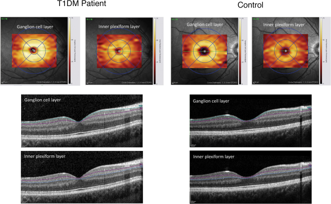
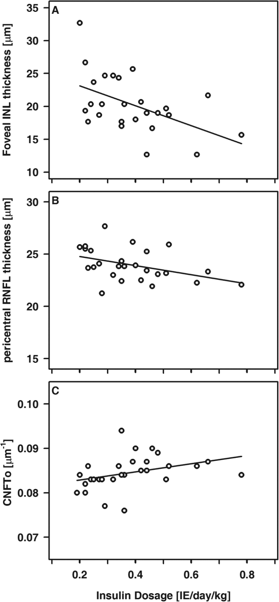
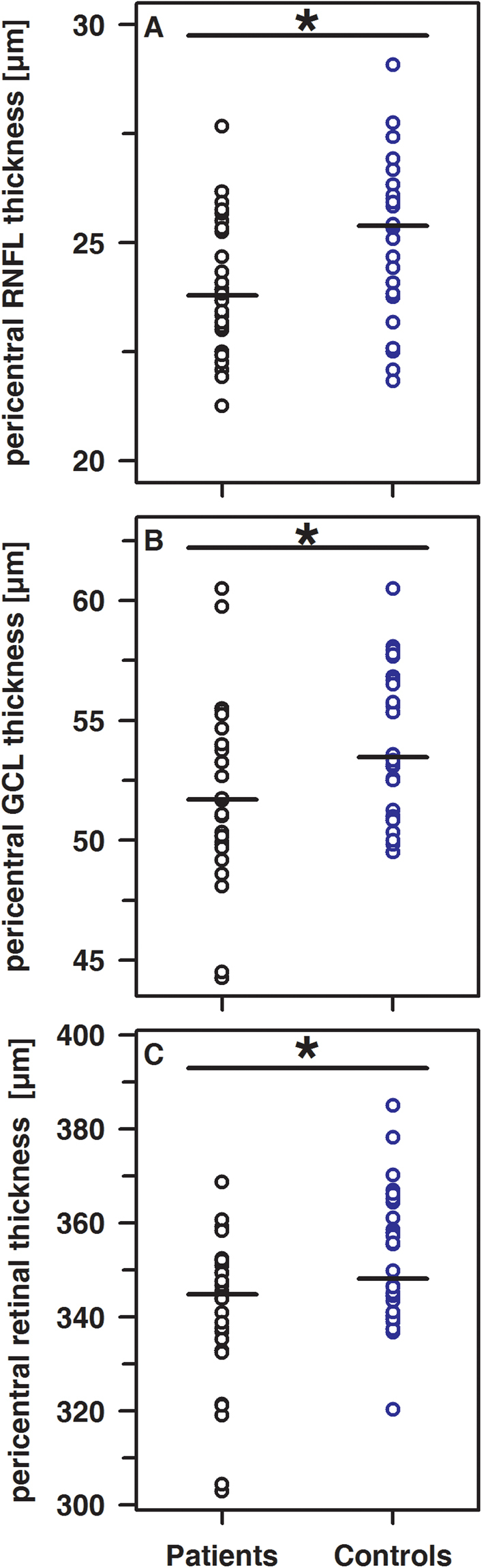
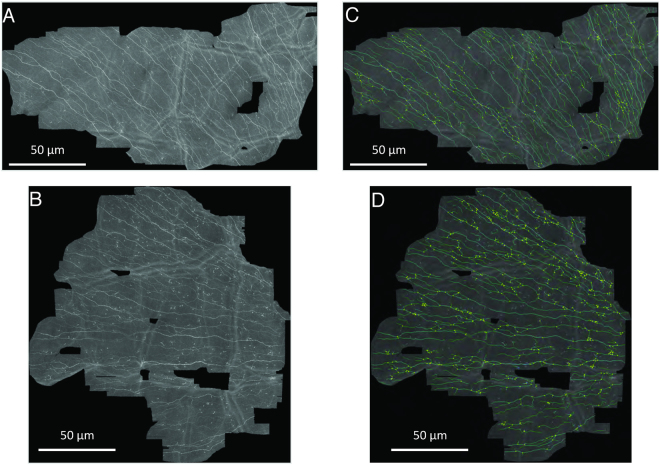
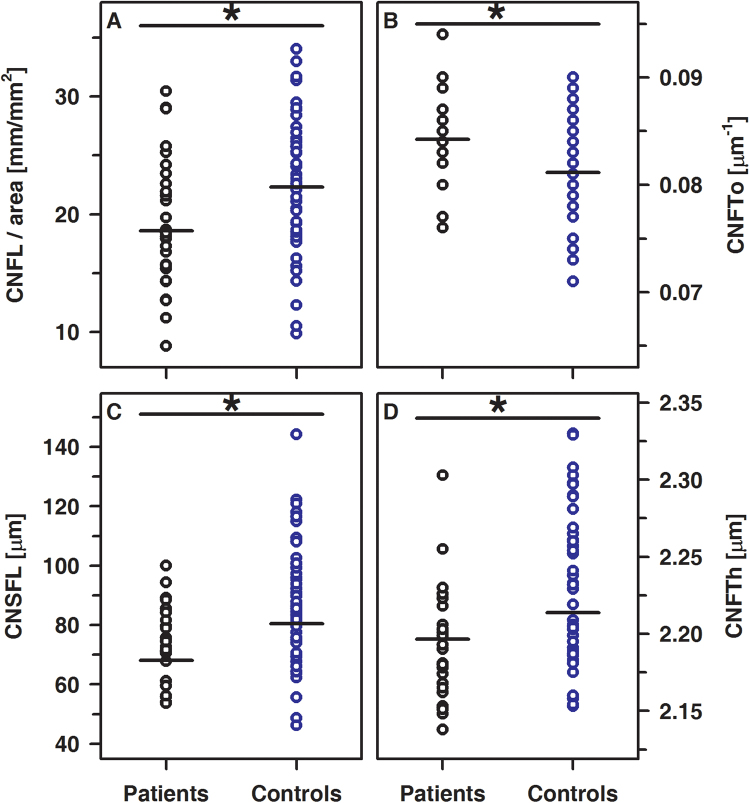
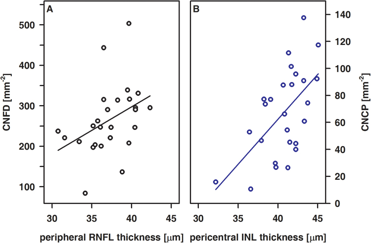
Optical coherence tomography (OCT) of the retina and corneal confocal laser scanning microscopy (CLSM) of the subbasal nerve plexus (SBP) are noninvasive techniques for quantification of the ocular neurodegenerative changes in individuals with type 1 diabetes mellitus (T1DM). In adult T1DM patients these changes are hardly related to T1DM only. Instead, ageing and/or lifestyle associated comorbidities have to be considered as putative confounding variables. Therefore, we investigated pediatric T1DM patients (n = 28; 14.2 ± 2.51 y; duration of disease: 5.39 ± 4.16 y) without clinical signs of diabetic retina disease, neuropathy, vasculopathy or nephropathy and compared our findings with those obtained in healthy controls (n = 46; 14.8 ± 1.89 y). The SBP was characterized by the averaged length, thickness, and tortuosity of nerve fibers as well as the number of branching and connecting points. OCT was used to determine the total thickness of the retina (ALL) and the thickness of each retinal layer. Both methods revealed signs of early neurodegenerative changes, e.g. thinning of distinct retinal layers at the pericentral ring and shortening of corneal nerve fibers that are already present in pediatric T1DM patients. Standardization of instruments and algorithms are urgently required to enable uniform comparison between different groups and define normative values to introduce in the clinical setting.

摘要

视网膜光学相干断层扫描(OCT)和角膜共焦激光扫描显微镜(CLSM)可用于对 1 型糖尿病(T1DM)患者的眼神经退行性变进行定量分析,这两种方法均为非侵入性技术。在成年 T1DM 患者中,这些变化与 T1DM 密切相关。相反,需要考虑与年龄相关的生活方式相关的合并症作为潜在的混杂变量。因此,我们研究了没有糖尿病视网膜病变、神经病变、血管病变或肾病临床症状的儿科 T1DM 患者(n = 28;14.2 ± 2.51 y;疾病持续时间:5.39 ± 4.16 y),并将我们的发现与健康对照组(n = 46;14.8 ± 1.89 y)进行了比较。通过纤维的平均长度、厚度和扭曲度以及分支和连接点的数量来描述 SBP。OCT 用于确定视网膜的总厚度(ALL)和每个视网膜层的厚度。这两种方法都显示出早期神经退行性变的迹象,例如在中心周围环处的特定视网膜层变薄和角膜神经纤维缩短,这些变化在儿科 T1DM 患者中已经存在。迫切需要对仪器和算法进行标准化,以便能够在不同组之间进行统一比较,并定义在临床环境中引入的正常值。

https://cdn.ncbi.nlm.nih.gov/pmc/blobs/66a2/5758564/c70c3615ed00/41598_2017_18284_Fig6_HTML.jpg
https://cdn.ncbi.nlm.nih.gov/pmc/blobs/66a2/5758564/9cfef5e720d7/41598_2017_18284_Fig1_HTML.jpg
https://cdn.ncbi.nlm.nih.gov/pmc/blobs/66a2/5758564/61bb07aace98/41598_2017_18284_Fig2_HTML.jpg
https://cdn.ncbi.nlm.nih.gov/pmc/blobs/66a2/5758564/99826841f396/41598_2017_18284_Fig3_HTML.jpg
https://cdn.ncbi.nlm.nih.gov/pmc/blobs/66a2/5758564/7f02aa9d3e79/41598_2017_18284_Fig4_HTML.jpg
https://cdn.ncbi.nlm.nih.gov/pmc/blobs/66a2/5758564/bf32ffd575ce/41598_2017_18284_Fig5_HTML.jpg
https://cdn.ncbi.nlm.nih.gov/pmc/blobs/66a2/5758564/c70c3615ed00/41598_2017_18284_Fig6_HTML.jpg

相似文献

1
The corneal subbasal nerve plexus and thickness of the retinal layers in pediatric type 1 diabetes and matched controls.儿童 1 型糖尿病患者与匹配对照者的角膜基底部神经丛和视网膜层厚度。
Sci Rep. 2018 Jan 8;8(1):14. doi: 10.1038/s41598-017-18284-z.
2
New ophthalmologic imaging techniques for detection and monitoring of neurodegenerative changes in diabetes: a systematic review.用于检测和监测糖尿病神经退行性病变的新型眼科影像学技术:系统评价。
Lancet Diabetes Endocrinol. 2015 Aug;3(8):653-63. doi: 10.1016/S2213-8587(15)00136-9. Epub 2015 Jul 13.
3
Neurodegeneration of the cornea and retina in patients with type 1 diabetes without clinical evidence of diabetic retinopathy.1 型糖尿病患者的角膜和视网膜神经退行性变,尽管无临床证据表明存在糖尿病性视网膜病变。
Front Endocrinol (Lausanne). 2022 Oct 5;13:790255. doi: 10.3389/fendo.2022.790255. eCollection 2022.
4
Imaging of the Corneal Subbasal Whorl-like Nerve Plexus: More Accurate Depiction of the Extent of Corneal Nerve Damage in Patients With Diabetes.角膜基底膜下涡状神经丛成像:更准确描绘糖尿病患者角膜神经损伤程度
Invest Ophthalmol Vis Sci. 2015 Aug;56(9):5417-23. doi: 10.1167/iovs.15-16609.
5
Longitudinal Changes in Corneal Cell and Nerve Fiber Morphology in Young Patients with Type 1 Diabetes with and without Diabetic Retinopathy: A 2-Year Follow-up Study.1 型糖尿病伴或不伴糖尿病视网膜病变年轻患者角膜细胞和神经纤维形态的纵向变化:一项为期 2 年的随访研究。
Invest Ophthalmol Vis Sci. 2019 Feb 1;60(2):830-837. doi: 10.1167/iovs.18-24516.
6
Corneal and Retinal Neuronal Degeneration in Early Stages of Diabetic Retinopathy.糖尿病视网膜病变早期的角膜和视网膜神经元变性
Invest Ophthalmol Vis Sci. 2017 Dec 1;58(14):6365-6373. doi: 10.1167/iovs.17-22736.
7
Can optical coherence tomography predict early retinal microvascular pathology in type 1 diabetic adolescents without minimal diabetic retinopathy? A single-centre study.光学相干断层扫描能否预测1型糖尿病青少年早期视网膜微血管病变而无轻度糖尿病视网膜病变?一项单中心研究。
J Pediatr Endocrinol Metab. 2015 Jan;28(1-2):139-46. doi: 10.1515/jpem-2013-0499.
8
Early Corneal Cellular and Nerve Fiber Pathology in Young Patients With Type 1 Diabetes Mellitus Identified Using Corneal Confocal Microscopy.使用角膜共焦显微镜在1型糖尿病年轻患者中发现的早期角膜细胞和神经纤维病理学
Invest Ophthalmol Vis Sci. 2016 Mar;57(3):853-8. doi: 10.1167/iovs.15-18735.
9
Decreased Corneal Sensation and Subbasal Nerve Density, and Thinned Corneal Epithelium as a Result of 360-Degree Laser Retinopexy.360 度激光视网膜光凝术后角膜知觉下降、基底神经密度降低和角膜上皮变薄。
Ophthalmology. 2015 Oct;122(10):2095-102. doi: 10.1016/j.ophtha.2015.06.010. Epub 2015 Jul 16.
10
[Confocal microscope examination of the corneal nerve plexus as biomarker for systemic diseases : View from the corneal nerve plexus on diabetes mellitus disease].[共聚焦显微镜检查角膜神经丛作为全身性疾病的生物标志物:从角膜神经丛看糖尿病]
Ophthalmologe. 2017 Jul;114(7):592-600. doi: 10.1007/s00347-017-0480-4.

引用本文的文献

1
Methods for the evaluation of corneal nerve fibres in diabetes mellitus by in vivo confocal microscopy: a scoping review.通过体内共聚焦显微镜评估糖尿病患者角膜神经纤维的方法:一项范围综述
BMJ Open. 2025 Sep 4;15(9):e098294. doi: 10.1136/bmjopen-2024-098294.
2
RETINAL MICROVASCULOPATHY WITH DIFFERENT INSULIN INFUSION THERAPIES IN CHILDREN WITH TYPE 1 DIABETES MELLITUS WITHOUT CLINICAL DIABETIC RETINOPATHY.1型糖尿病无临床糖尿病视网膜病变患儿不同胰岛素输注疗法与视网膜微血管病变
Retina. 2024 May 1;44(5):895-900. doi: 10.1097/IAE.0000000000004028. Epub 2024 Apr 18.
3
Taxane-Induced Neuropathy and Its Ocular Effects-A Longitudinal Follow-up Study in Breast Cancer Patients.

本文引用的文献

1
A Novel Approach to Analyze the Progression of Measured Corneal Sub-Basal Nerve Fiber Length in Continuously Expanding Mosaic Images.一种分析连续扩展的镶嵌图像中测量的角膜基底膜下神经纤维长度进展的新方法。
Curr Eye Res. 2017 Apr;42(4):549-556. doi: 10.1080/02713683.2016.1221977. Epub 2016 Oct 21.
2
Retinal Nerve Fiber Layer and Ganglion Cell Complex Thicknesses Are Reduced in Children With Type 1 Diabetes With No Evidence of Vascular Retinopathy.1型糖尿病且无糖尿病视网膜病变证据的儿童视网膜神经纤维层和神经节细胞复合体厚度降低。
Invest Ophthalmol Vis Sci. 2016 Oct 1;57(13):5355-5360. doi: 10.1167/iovs.16-19988.
3
紫杉烷诱导的神经病变及其眼部影响——乳腺癌患者的纵向随访研究
Cancers (Basel). 2023 Apr 25;15(9):2444. doi: 10.3390/cancers15092444.
4
S1P promotes corneal trigeminal neuron differentiation and corneal nerve repair via upregulating nerve growth factor expression in a mouse model.在小鼠模型中,鞘氨醇-1-磷酸(S1P)通过上调神经生长因子表达促进角膜三叉神经节神经元分化和角膜神经修复。
Open Life Sci. 2022 Oct 12;17(1):1324-1332. doi: 10.1515/biol-2022-0491. eCollection 2022.
5
Attenuation of inflammation in streptozotocin-induced diabetic rabbits by oil: A focus on targeting NF-κB and NLRP3 signaling pathways.油对链脲佐菌素诱导的糖尿病兔炎症的减轻作用:聚焦于靶向核因子κB和NLRP3信号通路
Chin Herb Med. 2019 Dec 24;12(1):73-78. doi: 10.1016/j.chmed.2019.12.003. eCollection 2020 Jan.
6
Corneal Confocal Microscopy as a Quantitative Imaging Biomarker of Diabetic Peripheral Neuropathy: A Review.角膜共聚焦显微镜作为糖尿病周围神经病变的定量成像生物标志物:综述
J Clin Med. 2022 Aug 31;11(17):5130. doi: 10.3390/jcm11175130.
7
A Cross-Sectional Study of Ocular Changes in Children and Adolescents with Diabetes Mellitus in Selected Health Facilities in Ghana.加纳部分选定医疗机构中糖尿病患儿和青少年眼部变化的横断面研究。
Int J Environ Res Public Health. 2022 Apr 27;19(9):5295. doi: 10.3390/ijerph19095295.
8
In vivo confocal microscopy study of corneal nerve alterations in children and youths with Type 1 diabetes.1 型糖尿病患儿和青少年角膜神经改变的活体共聚焦显微镜研究。
Pediatr Diabetes. 2021 Aug;22(5):780-786. doi: 10.1111/pedi.13219. Epub 2021 May 11.
9
Early corneal nerve fibre damage and increased Langerhans cell density in children with type 1 diabetes mellitus.儿童 1 型糖尿病患者早期角膜神经纤维损伤和朗格汉斯细胞密度增加。
Sci Rep. 2019 Jun 19;9(1):8758. doi: 10.1038/s41598-019-45116-z.
10
Diagnosing Diabetic Neuropathy: Something Old, Something New.诊断糖尿病性神经病变:旧法新用
Diabetes Metab J. 2018 Aug;42(4):255-269. doi: 10.4093/dmj.2018.0056.
Is the incidence of type 1 diabetes in children and adolescents stabilising? The first 6 years of a National Register.
儿童和青少年1型糖尿病的发病率是否趋于稳定?国家登记处的头6年情况。
Eur J Pediatr. 2016 Dec;175(12):1913-1919. doi: 10.1007/s00431-016-2787-6. Epub 2016 Sep 22.
4
Evaluation of the correlation between type 1 diabetes and cognitive function in children and adolescents, and comparison of this correlation with structural changes in the central nervous system: a study protocol.评估 1 型糖尿病与儿童和青少年认知功能的相关性,并与中枢神经系统结构变化进行比较:研究方案。
BMJ Open. 2016 Apr 28;6(4):e007917. doi: 10.1136/bmjopen-2015-007917.
5
Diabetic macular morphology changes may occur in the early stage of diabetes.糖尿病性黄斑形态改变可能在糖尿病早期就会出现。
BMC Ophthalmol. 2016 Jan 18;16:12. doi: 10.1186/s12886-016-0186-4.
6
In Vivo Confocal Microscopy of the Human Cornea in the Assessment of Peripheral Neuropathy and Systemic Diseases.人体角膜的体内共聚焦显微镜检查在评估周围神经病变和全身性疾病中的应用
Biomed Res Int. 2015;2015:951081. doi: 10.1155/2015/951081. Epub 2015 Dec 7.
7
Structural Changes by Spectral Domain Optical Coherence Tomography in Patients With Type 1 Diabetes Mellitus.1型糖尿病患者经光谱域光学相干断层扫描的结构变化
J Diabetes Sci Technol. 2015 Aug 31;10(2):271-6. doi: 10.1177/1932296815603371.
8
New ophthalmologic imaging techniques for detection and monitoring of neurodegenerative changes in diabetes: a systematic review.用于检测和监测糖尿病神经退行性病变的新型眼科影像学技术:系统评价。
Lancet Diabetes Endocrinol. 2015 Aug;3(8):653-63. doi: 10.1016/S2213-8587(15)00136-9. Epub 2015 Jul 13.
9
Local Variability of Parameters for Characterization of the Corneal Subbasal Nerve Plexus.用于表征角膜基底神经丛的参数的局部变异性
Curr Eye Res. 2016;41(2):186-98. doi: 10.3109/02713683.2015.1010686. Epub 2015 Mar 24.
10
Normative values for corneal nerve morphology assessed using corneal confocal microscopy: a multinational normative data set.使用角膜共焦显微镜评估的角膜神经形态学的标准值:一个跨国标准数据集。
Diabetes Care. 2015 May;38(5):838-43. doi: 10.2337/dc14-2311. Epub 2015 Jan 29.