Cosmo Eleonora, Midena Giulia, Frizziero Luisa, Bruno Marisa, Cecere Michela, Midena Edoardo
IRCCS-Fondazione Bietti, 00198 Rome, Italy.
Department of Neuroscience-Ophthalmology, University of Padova, 35128 Padova, Italy.
J Clin Med. 2022 Aug 31;11(17):5130. doi: 10.3390/jcm11175130.
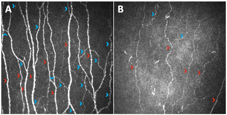
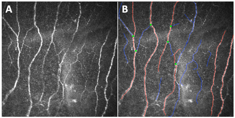
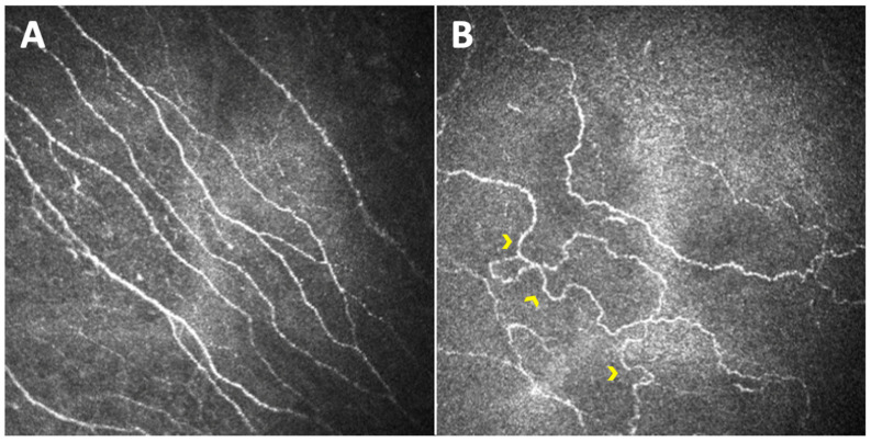
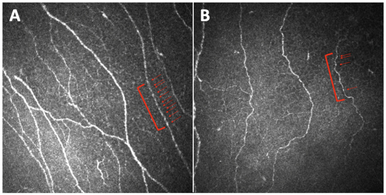
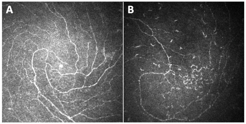
Distal symmetric polyneuropathy (DPN), particularly chronic sensorimotor DPN, represents one of the most frequent complications of diabetes, affecting 50% of diabetic patients and causing an enormous financial burden. Whilst diagnostic methods exist to detect and monitor this condition, they have significant limitations, mainly due to their high subjectivity, invasiveness, and non-repeatability. Corneal confocal microscopy (CCM) is an in vivo, non-invasive, and reproducible diagnostic technique for the study of all corneal layers including the sub-basal nerve plexus, which represents part of the peripheral nervous system. We reviewed the current literature on the use of CCM as an instrument in the assessment of diabetic patients, particularly focusing on its role in the study of sub-basal nerve plexus alterations as a marker of DPN. CCM has been demonstrated to be a valid in vivo tool to detect early sub-basal nerve plexus damage in adult and pediatric diabetic patients, correlating with the severity of DPN. Despite its great potential, CCM has still limited application in daily clinical practice, and more efforts still need to be made to allow the dissemination of this technique among doctors taking care of diabetic patients.
远端对称性多发性神经病(DPN),尤其是慢性感觉运动性DPN,是糖尿病最常见的并发症之一,影响着50%的糖尿病患者,并造成巨大的经济负担。虽然存在检测和监测这种疾病的诊断方法,但它们有显著局限性,主要是因为其主观性强、具有侵入性且不可重复。角膜共焦显微镜检查(CCM)是一种用于研究包括基底膜下神经丛在内的所有角膜层的体内、非侵入性且可重复的诊断技术,而基底膜下神经丛是周围神经系统的一部分。我们回顾了有关将CCM用作评估糖尿病患者的工具的当前文献,特别关注其在研究基底膜下神经丛改变作为DPN标志物方面的作用。CCM已被证明是一种有效的体内工具,可检测成人和儿童糖尿病患者早期基底膜下神经丛损伤,且与DPN的严重程度相关。尽管CCM潜力巨大,但在日常临床实践中的应用仍然有限,仍需要做出更多努力,以使这项技术在照顾糖尿病患者的医生中得到推广。