Department of Ophthalmology and Visual Science, College of Medicine, The Catholic University of Korea, Seoul, Republic of Korea.
Catholic Institute for Visual Science, College of Medicine, The Catholic University of Korea, Seoul, Korea.
Sci Rep. 2018 Jan 8;8(1):70. doi: 10.1038/s41598-017-18511-7.
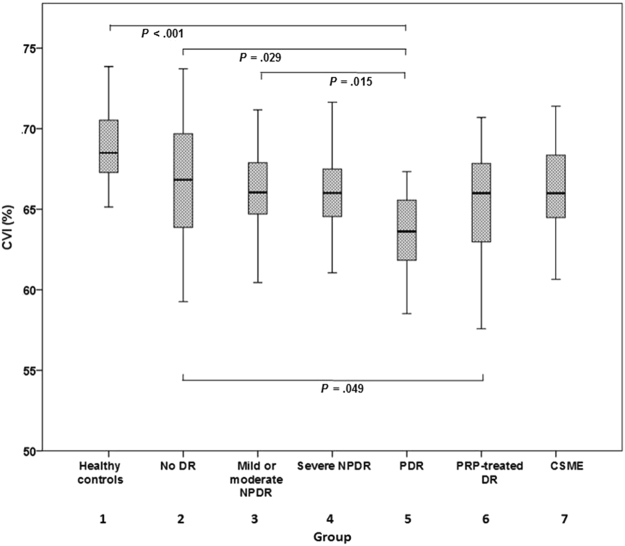
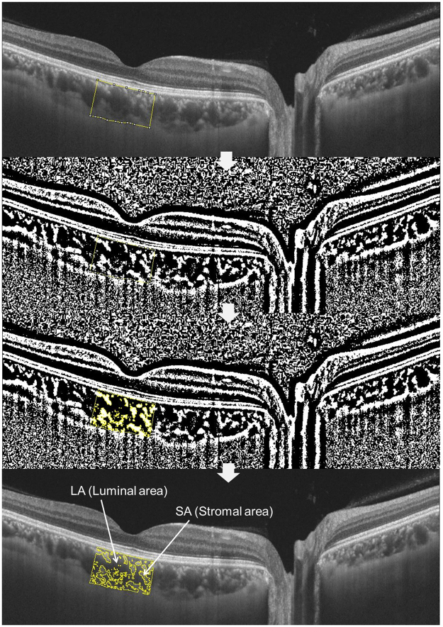
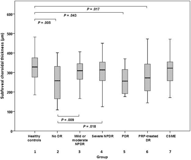
The relationships between changes in choroidal vasculature and the severity of diabetic retinopathy (DR) remain unclear. We assessed choroidal changes in diabetic patients by measuring choroidal vascularity index (CVI) in conjunction with DR stage. In this study, patients with diabetes and healthy controls were retrospectively analyzed. Subjects were divided into seven groups as follows: Healthy controls, no DR, mild/moderate non-proliferative DR (NPDR), severe NPDR, proliferative DR (PDR), panretinal photocoagulation-treated DR, and clinically significant macular edema. The mean CVI values in the above groups were 69.08, 67.07, 66.28, 66.20, 63.48, 65.38, and 66.28, respectively. The eyes of diabetic patients exhibited a significantly lower CVI value than those of healthy controls even without DR. The PDR group exhibited a significantly lower CVI value than the healthy control, no DR, and mild/moderate NPDR groups. Age, sex, disease duration, glycated hemoglobin, fasting blood sugar, or intraocular pressure had no correlation with CVI. In multivariate regression analysis, thicker subfoveal choroid and thinner central retina were significantly associated with higher CVI values. These findings carefully suggest that changes in choroidal vasculature could be the primary event in diabetes even where there is no DR.
脉络膜血管变化与糖尿病视网膜病变(DR)严重程度之间的关系尚不清楚。我们通过测量脉络膜血管密度指数(CVI)并结合 DR 分期来评估糖尿病患者的脉络膜变化。在这项研究中,回顾性分析了糖尿病患者和健康对照者。受试者分为以下七组:健康对照组、无 DR、轻度/中度非增殖性 DR(NPDR)、重度 NPDR、增殖性 DR(PDR)、全视网膜光凝治疗 DR 和临床显著黄斑水肿。上述各组的平均 CVI 值分别为 69.08、67.07、66.28、66.20、63.48、65.38 和 66.28。即使没有 DR,糖尿病患者的眼睛也表现出明显较低的 CVI 值。PDR 组的 CVI 值明显低于健康对照组、无 DR 组和轻度/中度 NPDR 组。年龄、性别、病程、糖化血红蛋白、空腹血糖或眼内压与 CVI 均无相关性。多元回归分析显示,脉络膜下较厚和中心视网膜较薄与较高的 CVI 值显著相关。这些发现提示,脉络膜血管变化可能是糖尿病的初始事件,即使没有 DR 也是如此。