• 文献检索
  • 文档翻译
  • 深度研究
  • 学术资讯
  • Suppr Zotero 插件Zotero 插件
  • 邀请有礼
  • 套餐&价格
  • 历史记录
应用&插件
Suppr Zotero 插件Zotero 插件浏览器插件Mac 客户端Windows 客户端微信小程序
定价
高级版会员购买积分包购买API积分包
服务
文献检索文档翻译深度研究API 文档MCP 服务
关于我们
关于 Suppr公司介绍联系我们用户协议隐私条款
关注我们

Suppr 超能文献

核心技术专利:CN118964589B侵权必究
粤ICP备2023148730 号-1Suppr @ 2026

文献检索

告别复杂PubMed语法,用中文像聊天一样搜索,搜遍4000万医学文献。AI智能推荐,让科研检索更轻松。

立即免费搜索

文件翻译

保留排版,准确专业,支持PDF/Word/PPT等文件格式,支持 12+语言互译。

免费翻译文档

深度研究

AI帮你快速写综述,25分钟生成高质量综述,智能提取关键信息,辅助科研写作。

立即免费体验

糖尿病视网膜病变中脉络膜血管变化及其与视力的相关性

Vascular changes of the choroid and their correlations with visual acuity in diabetic retinopathy.

作者信息

Jing Ruixia, Sun Xiubin, Cheng Jimin, Li Xue, Wang Zhen

机构信息

Jinan Central Hospital, Shandong First Medical University & Shandong Academy of Medical Sciences, Jinan, China.

Department of Biostatistics, School of Public Health, Cheeloo Collage of Medicine, Shandong University, Jinan, China.

出版信息

Front Endocrinol (Lausanne). 2024 Feb 23;15:1327325. doi: 10.3389/fendo.2024.1327325. eCollection 2024.

DOI:10.3389/fendo.2024.1327325
PMID:38464970
原文链接:https://pmc.ncbi.nlm.nih.gov/articles/PMC10920230/
Abstract

OBJECTIVE

To investigate changes in the choroidal vasculature and their correlations with visual acuity in diabetic retinopathy (DR).

METHODS

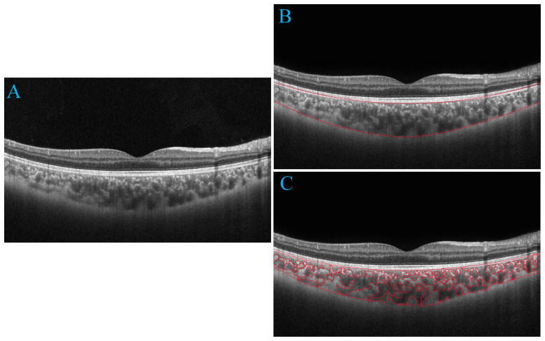
The cohort was composed of 225 eyes from 225 subjects, including 60 eyes from 60 subjects with healthy control, 55 eyes from 55 subjects without DR, 46 eyes from 46 subjects with nonproliferative diabetic retinopathy (NPDR), 21 eyes from 21 subjects with proliferative diabetic retinopathy (PDR), and 43 eyes from 43 subjects with clinically significant macular edema (CSME). Swept-source optical coherence tomography (SS-OCT) was used to image the eyes with a 12-mm radial line scan protocol. The parameters for 6-mm diameters of region centered on the macular fovea were analyzed. Initially, a custom deep learning algorithm based on a modified residual U-Net architecture was utilized for choroidal boundary segmentation. Subsequently, the SS-OCT image was binarized and the Niblack-based automatic local threshold algorithm was employed to calibrate subfoveal choroidal thickness (SFCT), luminal area (LA), and stromal area (SA) by determining the distance between the two boundaries. Finally, the ratio of LA and total choroidal area (SA + LA) was defined as the choroidal vascularity index (CVI). The choroidal parameters in five groups were compared, and correlations of the choroidal parameters with age, gender, duration of diabetes mellitus (DM), glycated hemoglobin (HbA1c), fasting blood sugar, SFCT and best-corrected visual acuity (BCVA) were analyzed.

RESULTS

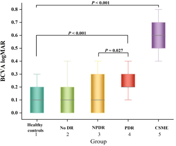
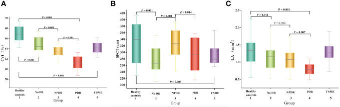
The CVI, SFCT, LA, and SA values of patients with DR were found to be significantly lower compared to both healthy patients and patients without DR ( < 0.05). The SFCT was significantly higher in NPDR group compared to the No DR group ( < 0.001). Additionally, the SFCT was lower in the PDR group compared to the NPDR group ( = 0.014). Furthermore, there was a gradual decrease in CVI with progression of diabetic retinopathy, reaching its lowest value in the PDR group. However, the CVI of the CSME group exhibited a marginally closer proximity to that of the NPDR group. The multivariate regression analysis revealed a positive correlation between CVI and the duration of DM as well as LA ( < 0.05). The results of both univariate and multivariate regression analyses demonstrated a significant positive correlation between CVI and BCVA ( = 0.003).

CONCLUSION

Choroidal vascular alterations, especially decreased CVI, occurred in patients with DR. The CVI decreased with duration of DM and was correlated with visual impairment, indicating that the CVI might be a reliable imaging biomarker to monitor the progression of DR.

摘要

目的

研究糖尿病视网膜病变(DR)患者脉络膜血管的变化及其与视力的相关性。

方法

该队列由225名受试者的225只眼睛组成,包括60名健康对照受试者的60只眼睛、55名无DR受试者的55只眼睛、46名非增殖性糖尿病视网膜病变(NPDR)受试者的46只眼睛、21名增殖性糖尿病视网膜病变(PDR)受试者的21只眼睛以及43名具有临床显著性黄斑水肿(CSME)受试者的43只眼睛。使用扫频光学相干断层扫描(SS-OCT)以12毫米径向线扫描方案对眼睛进行成像。分析以黄斑中心凹为中心的直径6毫米区域的参数。首先,利用基于改进残差U-Net架构的定制深度学习算法进行脉络膜边界分割。随后,将SS-OCT图像二值化,并采用基于尼布莱克的自动局部阈值算法,通过确定两个边界之间的距离来校准中心凹下脉络膜厚度(SFCT)、管腔面积(LA)和基质面积(SA)。最后,将LA与脉络膜总面积(SA+LA)的比值定义为脉络膜血管指数(CVI)。比较五组的脉络膜参数,并分析脉络膜参数与年龄、性别、糖尿病病程(DM)、糖化血红蛋白(HbA1c)、空腹血糖、SFCT和最佳矫正视力(BCVA)的相关性。

结果

发现DR患者的CVI、SFCT、LA和SA值与健康患者和无DR患者相比均显著降低(P<0.05)。NPDR组的SFCT显著高于无DR组(P<0.001)。此外,PDR组的SFCT低于NPDR组(P=0.014)。此外,随着糖尿病视网膜病变的进展,CVI逐渐降低,在PDR组达到最低值。然而,CSME组的CVI与NPDR组的CVI略有接近。多因素回归分析显示CVI与DM病程以及LA呈正相关(P<0.05)。单因素和多因素回归分析结果均显示CVI与BCVA呈显著正相关(P=0.003)。

结论

DR患者发生脉络膜血管改变,尤其是CVI降低。CVI随DM病程降低,并与视力损害相关,表明CVI可能是监测DR进展的可靠影像学生物标志物。

https://cdn.ncbi.nlm.nih.gov/pmc/blobs/450f/10920230/e9b91fc80c9e/fendo-15-1327325-g003.jpg
https://cdn.ncbi.nlm.nih.gov/pmc/blobs/450f/10920230/0c1da2b70a7d/fendo-15-1327325-g001.jpg
https://cdn.ncbi.nlm.nih.gov/pmc/blobs/450f/10920230/541b65a705e4/fendo-15-1327325-g002.jpg
https://cdn.ncbi.nlm.nih.gov/pmc/blobs/450f/10920230/e9b91fc80c9e/fendo-15-1327325-g003.jpg
https://cdn.ncbi.nlm.nih.gov/pmc/blobs/450f/10920230/0c1da2b70a7d/fendo-15-1327325-g001.jpg
https://cdn.ncbi.nlm.nih.gov/pmc/blobs/450f/10920230/541b65a705e4/fendo-15-1327325-g002.jpg
https://cdn.ncbi.nlm.nih.gov/pmc/blobs/450f/10920230/e9b91fc80c9e/fendo-15-1327325-g003.jpg

相似文献

1
Vascular changes of the choroid and their correlations with visual acuity in diabetic retinopathy.糖尿病视网膜病变中脉络膜血管变化及其与视力的相关性
Front Endocrinol (Lausanne). 2024 Feb 23;15:1327325. doi: 10.3389/fendo.2024.1327325. eCollection 2024.
2
Choroidal structural analysis in eyes with diabetic retinopathy and diabetic macular edema-A novel OCT based imaging biomarker.糖尿病性视网膜病变和糖尿病性黄斑水肿患者脉络膜结构分析 - 一种新型基于 OCT 的成像生物标志物。
PLoS One. 2018 Dec 11;13(12):e0207435. doi: 10.1371/journal.pone.0207435. eCollection 2018.
3
Choroidal Assessment in Patients with Type 2 Diabetes Mellitus and Non-Proliferative Diabetic Retinopathy by Swept-Source Ocular Coherence Tomography and Image Binarization.应用扫频源光相干断层扫描和图像二值化技术评估 2 型糖尿病非增殖性糖尿病视网膜病变患者脉络膜
Medicina (Kaunas). 2022 Jul 10;58(7):918. doi: 10.3390/medicina58070918.
4
Vascular Changes of the Choroid and Their Correlations With Visual Acuity in Pathological Myopia.病理性近视脉络膜血管改变及其与视力的相关性。
Invest Ophthalmol Vis Sci. 2022 Nov 1;63(12):20. doi: 10.1167/iovs.63.12.20.
5
[Association of choroidal thickness with diabetic retinopathy at different stages].[不同阶段糖尿病视网膜病变与脉络膜厚度的相关性]
Zhonghua Yi Xue Za Zhi. 2015 Aug 25;95(32):2584-8.
6
Choroidal vascularity index (CVI)-Net-based automatic assessment of diabetic retinopathy severity using CVI in optical coherence tomography images.基于脉络膜血管密度指数(CVI)网络的光学相干断层扫描图像中 CVI 自动评估糖尿病视网膜病变严重程度。
J Biophotonics. 2023 Jun;16(6):e202200370. doi: 10.1002/jbio.202200370. Epub 2023 Feb 16.
7
The quantitative measurements of choroidal thickness and volume in diabetic retinopathy using optical coherence tomography and optical coherence tomography angiography; correlation with vision and foveal avascular zone.应用光学相干断层扫描和光学相干断层扫描血管成像对糖尿病视网膜病变脉络膜厚度和体积的定量测量;与视力和中心无血管区的相关性。
BMC Ophthalmol. 2022 Jan 3;22(1):3. doi: 10.1186/s12886-021-02178-w.
8
Choroidal structural changes correlate with severity of diabetic retinopathy in diabetes mellitus.脉络膜结构变化与糖尿病性视网膜病变的严重程度相关。
BMC Ophthalmol. 2019 Aug 16;19(1):186. doi: 10.1186/s12886-019-1189-8.
9
Alterations in the Choroidal Sublayers in Relationship to Severity and Progression of Diabetic Retinopathy: A Swept-Source OCT Study.与糖尿病视网膜病变严重程度和进展相关的脉络膜各层改变:一项扫频光学相干断层扫描研究
Ophthalmol Sci. 2022 Feb 25;2(2):100130. doi: 10.1016/j.xops.2022.100130. eCollection 2022 Jun.
10
Choroidal vascularity index in type-2 diabetes analyzed by swept-source optical coherence tomography.应用扫频源光相干断层扫描分析 2 型糖尿病脉络膜血流指数。
Sci Rep. 2018 Jan 8;8(1):70. doi: 10.1038/s41598-017-18511-7.

引用本文的文献

1
Choroidal vascularity index is independent of ocular and image-based factors in healthy eyes: a systematic review and meta-analysis.脉络膜血管指数在健康眼中独立于眼部及基于图像的因素:一项系统评价与荟萃分析
Sci Rep. 2025 Jul 30;15(1):27782. doi: 10.1038/s41598-025-10384-5.
2
Systemic inflammatory markers and neurovascular changes in the retina and choroid of diabetic patients without retinopathy: insights from wide-field SS-OCTA.无视网膜病变的糖尿病患者视网膜和脉络膜中的全身炎症标志物及神经血管变化:来自广角 SS-OCTA 的见解
Front Med (Lausanne). 2025 Jun 4;12:1566047. doi: 10.3389/fmed.2025.1566047. eCollection 2025.
3

本文引用的文献

1
Vascular Changes of the Choroid and Their Correlations With Visual Acuity in Pathological Myopia.病理性近视脉络膜血管改变及其与视力的相关性。
Invest Ophthalmol Vis Sci. 2022 Nov 1;63(12):20. doi: 10.1167/iovs.63.12.20.
2
Prophylactic interventions for preventing macular edema after cataract surgery in patients with diabetes: A Bayesian network meta-analysis of randomized controlled trials.糖尿病患者白内障手术后预防黄斑水肿的预防性干预措施:一项随机对照试验的贝叶斯网络荟萃分析。
EClinicalMedicine. 2022 May 20;49:101463. doi: 10.1016/j.eclinm.2022.101463. eCollection 2022 Jul.
3
Choroidal structural alterations in diabetic patients in association with disease duration, HbA1c level, and presence of retinopathy.
Three-Dimensional Choroidal Vessels Assessment in Diabetic Retinopathy.
糖尿病视网膜病变中脉络膜血管的三维评估
Invest Ophthalmol Vis Sci. 2025 Mar 3;66(3):50. doi: 10.1167/iovs.66.3.50.
糖尿病患者脉络膜结构改变与疾病持续时间、HbA1c 水平和视网膜病变的关系。
Int Ophthalmol. 2022 Dec;42(12):3661-3672. doi: 10.1007/s10792-022-02363-w. Epub 2022 May 23.
4
Macular Choroidal Thickness and the Risk of Referable Diabetic Retinopathy in Type 2 Diabetes: A 2-Year Longitudinal Study.黄斑脉络膜厚度与 2 型糖尿病可治疗性糖尿病视网膜病变风险:一项 2 年纵向研究。
Invest Ophthalmol Vis Sci. 2022 Apr 1;63(4):9. doi: 10.1167/iovs.63.4.9.
5
Retinal and Choroidal Vascular Perfusion and Thickness Measurement in Diabetic Retinopathy Patients by the Swept-Source Optical Coherence Tomography Angiography.通过扫频源光学相干断层扫描血管造影术测量糖尿病视网膜病变患者的视网膜和脉络膜血管灌注及厚度
Front Med (Lausanne). 2022 Mar 18;9:786708. doi: 10.3389/fmed.2022.786708. eCollection 2022.
6
Choroidal changes and associations with visual acuity in diabetic patients.糖尿病患者的脉络膜变化及其与视力的关系。
Int J Retina Vitreous. 2022 Jan 8;8(1):6. doi: 10.1186/s40942-021-00355-z.
7
Automatic Segmentation and Visualization of Choroid in OCT with Knowledge Infused Deep Learning.基于知识融合深度学习的 OCT 脉络膜自动分割与可视化。
IEEE J Biomed Health Inform. 2020 Dec;24(12):3408-3420. doi: 10.1109/JBHI.2020.3023144. Epub 2020 Dec 4.
8
Choroidal Thickness in Diabetes and Diabetic Retinopathy: A Swept Source OCT Study.脉络膜厚度在糖尿病和糖尿病性视网膜病变中的研究:扫频源光学相干断层扫描。
Invest Ophthalmol Vis Sci. 2020 Apr 9;61(4):29. doi: 10.1167/iovs.61.4.29.
9
Interpretation of OCT and OCTA images from a histological approach: Clinical and experimental implications.从组织学角度解读 OCT 和 OCTA 图像:临床和实验意义。
Prog Retin Eye Res. 2020 Jul;77:100828. doi: 10.1016/j.preteyeres.2019.100828. Epub 2020 Jan 3.
10
Mapping choroidal thickness in patients with type 2 diabetes.测量 2 型糖尿病患者脉络膜厚度。
Can J Ophthalmol. 2020 Feb;55(1):45-51. doi: 10.1016/j.jcjo.2019.06.009. Epub 2019 Aug 9.