Department of Pharmacology, Wayne State University School of Medicine, Detroit, MI, USA.
Karmanos Cancer Institute, Wayne State University School of Medicine, Detroit, MI, USA.
Sci Rep. 2018 Jan 8;8(1):40. doi: 10.1038/s41598-017-17800-5.
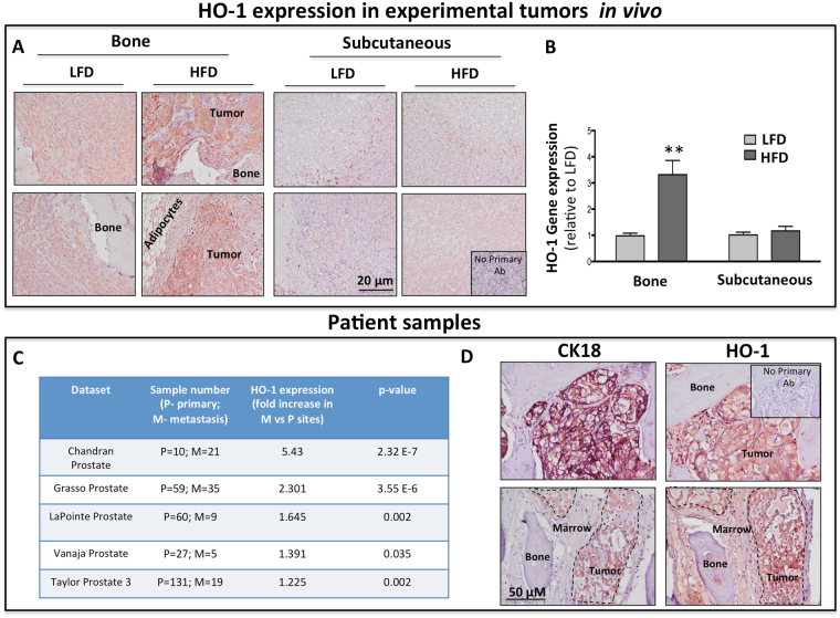
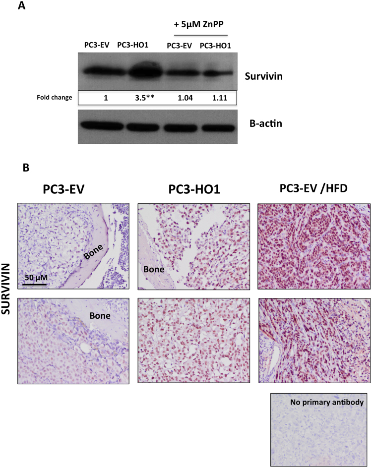
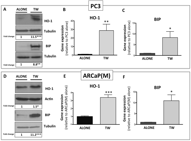
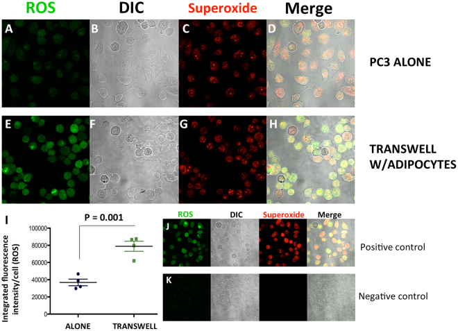
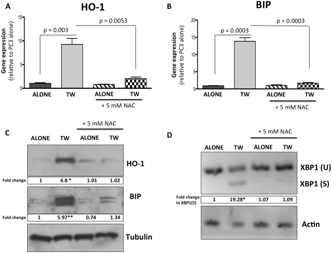
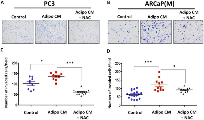
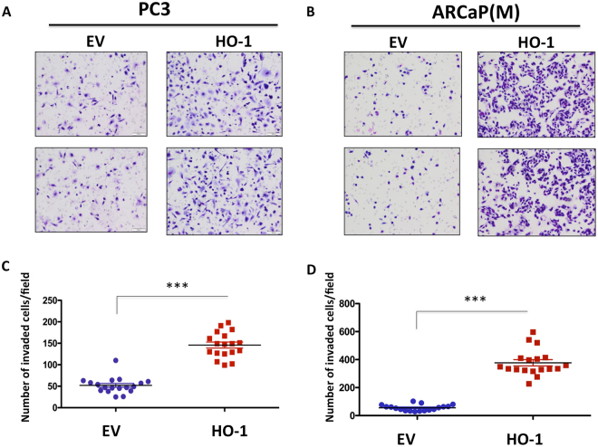
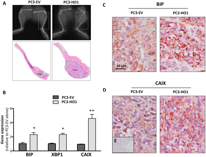
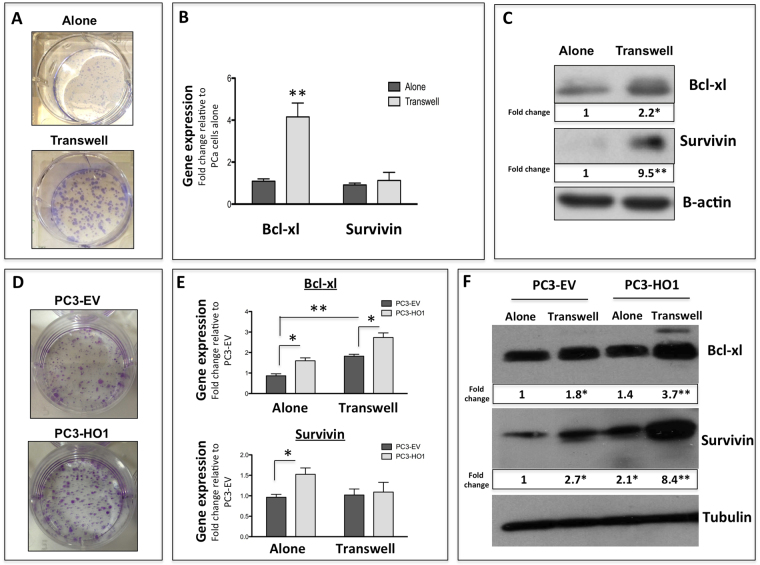
Metastatic tumor cells engage the local tumor microenvironment and activate specific pro-survival mechanisms to thrive and progress in the harsh bone marrow niche. Here we show that the major contributors to the survival of carcinoma cells that have colonized the bone marrow are the adipocyte-induced oxidative stress and ER stress pathways. We demonstrate that upon exposure to adipocyte-rich environments in vitro or in vivo, bone-trophic prostate and breast tumor cells upregulate the oxidative stress enzyme, HO-1. We also show that HO-1 levels are significantly increased in human metastatic prostate cancer tissues and that stable HO-1 overexpression in tumor cells promotes growth and invasiveness. Co-incident with the adipocyte-induced expression of HO-1, there is an upregulation of ER chaperone BIP and splicing of XBP1, indicating adipocyte-driven unfolded protein response, a process that we show to be sensitive to antioxidant treatment. Importantly, we also demonstrate that triggering of the oxidative stress and ER stress responses, or HO-1 induction by adipocyte exposure result in the activation of pro-survival pathways, involving survivin. Collectively, our findings reveal a new link between HO-1 and survivin expression in tumor cells, and provide a new insight into potentially targetable survival pathways in bone-metastatic disease.
转移瘤细胞与局部肿瘤微环境相互作用,并激活特定的生存促进机制,在恶劣的骨髓微环境中茁壮成长和发展。在这里,我们表明,导致已定植于骨髓中的癌细胞存活的主要因素是脂肪细胞诱导的氧化应激和内质网应激途径。我们证明,体外或体内暴露于富含脂肪细胞的环境中时,骨营养性前列腺癌和乳腺癌细胞会上调氧化应激酶 HO-1。我们还表明,人转移性前列腺癌组织中 HO-1 的水平显著增加,并且肿瘤细胞中 HO-1 的稳定过表达可促进其生长和侵袭性。与脂肪细胞诱导的 HO-1 表达同时发生的是内质网伴侣 BIP 和 XBP1 剪接的上调,表明脂肪细胞驱动的未折叠蛋白反应,我们发现该过程对抗氧化剂治疗敏感。重要的是,我们还证明,氧化应激和内质网应激反应的触发,或脂肪细胞暴露导致 HO-1 诱导,会激活涉及生存素的生存促进途径。总之,我们的发现揭示了肿瘤细胞中 HO-1 和生存素表达之间的新联系,并为骨转移疾病中潜在的可靶向生存途径提供了新的见解。