European Synchrotron Radiation Facility (ESRF), Grenoble, France.
Department of Biological Science and Program in Neuroscience, Florida State University, Tallahassee, FL, USA.
Sci Rep. 2018 Jan 9;8(1):184. doi: 10.1038/s41598-017-18000-x.
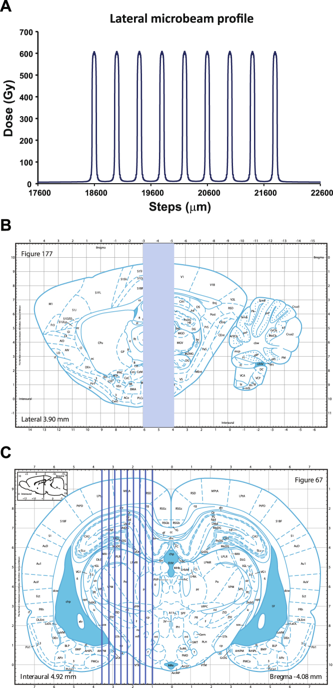
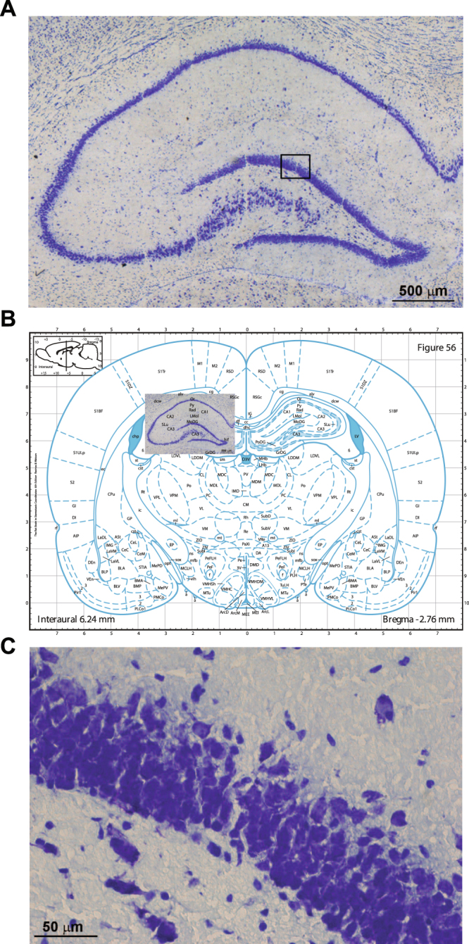
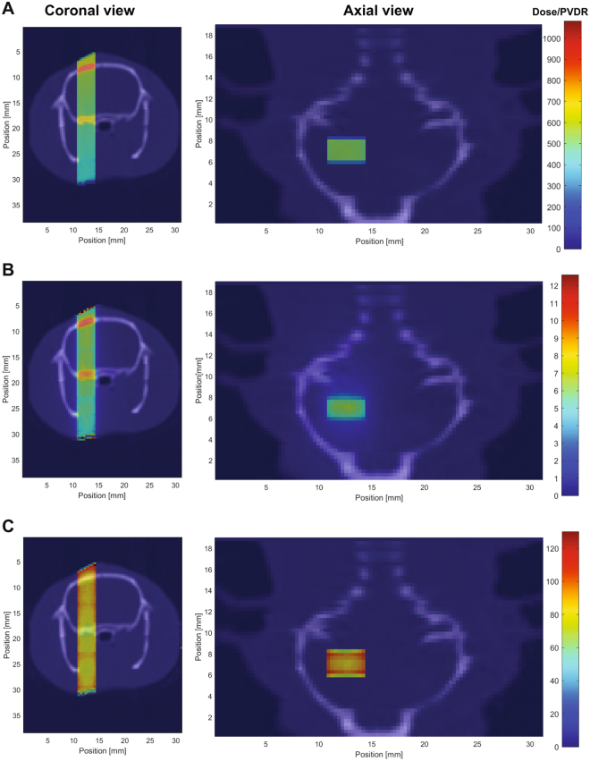
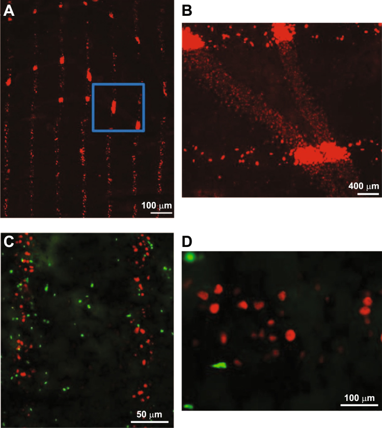
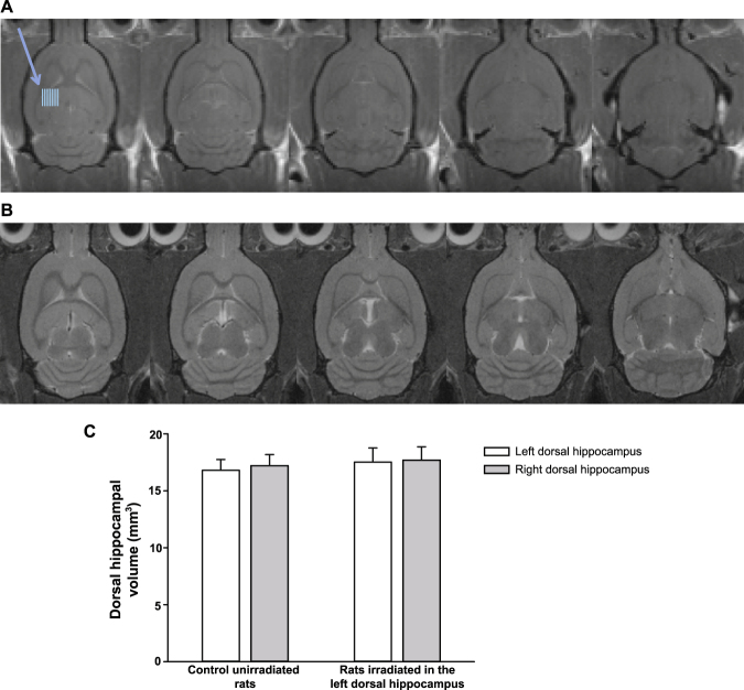
Synchrotron-generated microplanar beams (microbeams) provide the most stereo-selective irradiation modality known today. This novel irradiation modality has been shown to control seizures originating from eloquent cortex causing no neurological deficit in experimental animals. To test the hypothesis that application of microbeams in the hippocampus, the most common source of refractory seizures, is safe and does not induce severe side effects, we used microbeams to induce transections to the hippocampus of healthy rats. An array of parallel microbeams carrying an incident dose of 600 Gy was delivered to the rat hippocampus. Immunohistochemistry of phosphorylated γ-H2AX showed cell death along the microbeam irradiation paths in rats 48 hours after irradiation. No evident behavioral or neurological deficits were observed during the 3-month period of observation. MR imaging showed no signs of radio-induced edema or radionecrosis 3 months after irradiation. Histological analysis showed a very well preserved hippocampal cytoarchitecture and confirmed the presence of clear-cut microscopic transections across the hippocampus. These data support the use of synchrotron-generated microbeams as a novel tool to slice the hippocampus of living rats in a minimally invasive way, providing (i) a novel experimental model to study hippocampal function and (ii) a new treatment tool for patients affected by refractory epilepsy induced by mesial temporal sclerosis.
同步辐射微平板束(microbeams)提供了目前最具立体选择性的放射治疗方式。这种新型的放射治疗方式已被证明可以控制起源于优势脑区的癫痫发作,而不会在实验动物中引起神经功能缺陷。为了验证在海马体(最常见的难治性癫痫发作源)中应用微束是安全的,并且不会引起严重的副作用的假设,我们使用微束在健康大鼠的海马体中诱导横断。将具有 600Gy 入射剂量的一系列平行微束输送到大鼠的海马体。照射后 48 小时,磷酸化 γ-H2AX 的免疫组织化学显示细胞沿着微束照射路径死亡。在 3 个月的观察期间,没有观察到明显的行为或神经功能缺陷。磁共振成像显示照射后 3 个月没有放射性水肿或放射性坏死的迹象。组织学分析显示海马体的细胞结构得到了很好的保存,并证实了横跨海马体的明显的微观横断。这些数据支持使用同步辐射微束作为一种新的工具,以微创的方式切割活大鼠的海马体,为研究海马体功能提供了(i)一种新的实验模型,以及(ii)一种治疗内侧颞叶硬化引起的难治性癫痫患者的新治疗工具。