Wyatt-Johnson Season K, Herr Seth A, Brewster Amy L
Department of Psychological Sciences, College of Health and Human Sciences, Purdue University, West Lafayette, IN, United States.
Weldon School of Biomedical Engineering, West Lafayette, IN, United States.
Front Neurol. 2017 Dec 18;8:700. doi: 10.3389/fneur.2017.00700. eCollection 2017.
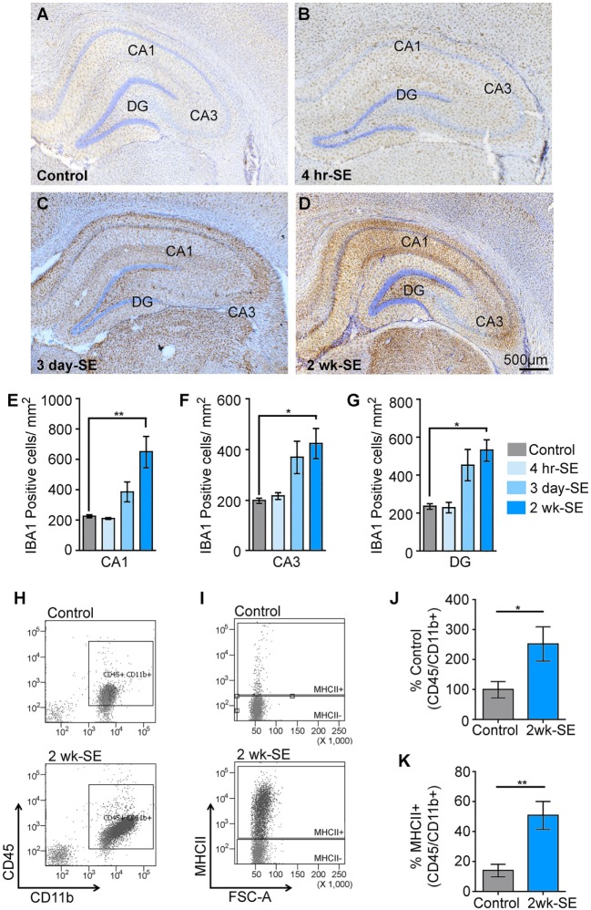
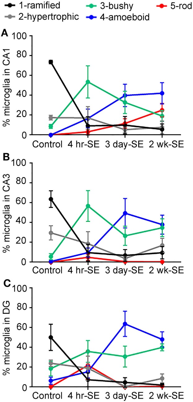
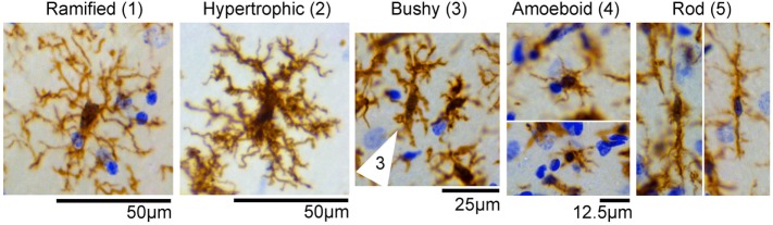
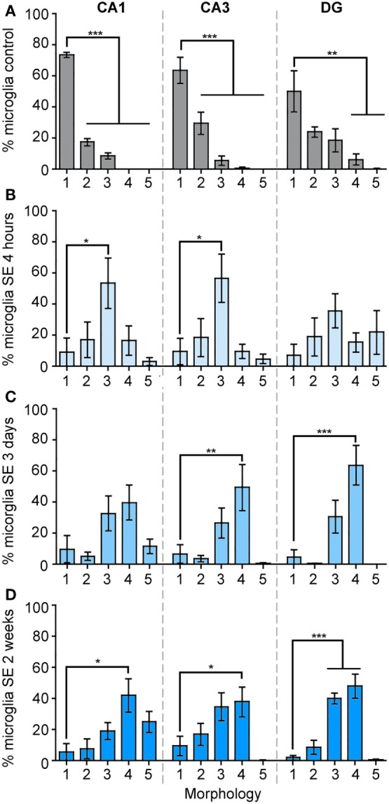
Status epilepticus (SE) is defined by the occurrence of prolonged "non-stop" seizures that last for at least 5 min. SE provokes inflammatory responses including the activation of microglial cells, the brain's resident immune cells, which are thought to contribute to the neuropathology and pathophysiology of epilepsy. Microglia are professional phagocytes that resemble peripheral macrophages. Upon sensing immune disturbances, including SE, microglia become reactive, produce inflammatory cytokines, and alter their actin cytoskeleton to transform from ramified to amoeboid shapes. It is widely known that SE triggers time-dependent microglial expression of pro-inflammatory cytokines that include TNFα and IL-1β. However, less is known in regards to the spatiotemporal progression of the morphological changes, which may help define the extent of microglia reactivity after SE and potential function (surveillance, inflammatory, phagocytic). Therefore, in this study, we used the microglia/macrophage IBA1 marker to identify and count these cells in hippocampi from control rats and at 4 h, 3 days, and 2 weeks after a single episode of pilocarpine-induced SE. We identified, categorized, and counted the IBA1-positive cells with the different morphologies observed after SE in the hippocampal areas CA1, CA3, and dentate gyrus. These included ramified, hypertrophic, bushy, amoeboid, and rod. We found that the ramified phenotype was the most abundant in control hippocampi. In contrast, SE provoked time-dependent changes in the microglial morphology that was characterized by significant increases in the abundance of bushy-shaped cells at 4 h and amoeboid-shaped cells at 3 days and 2 weeks. Interestingly, a significant increase in the number of rod-shaped cells was only evident in the CA1 region at 2 weeks after SE. Taken together, these data suggest that SE triggers time-dependent alterations in the morphology of microglial cells. This detailed description of the spatiotemporal profile of SE-induced microglial morphological changes may help provide insight into their contribution to epileptogenesis.
癫痫持续状态(SE)的定义是出现持续至少5分钟的长时间“不停歇”发作。SE会引发炎症反应,包括激活小胶质细胞,即大脑中的常驻免疫细胞,人们认为这些细胞会导致癫痫的神经病理学和病理生理学变化。小胶质细胞是类似于外周巨噬细胞的专业吞噬细胞。在感知包括SE在内的免疫干扰时,小胶质细胞会变得活跃,产生炎性细胞因子,并改变其肌动蛋白细胞骨架,从分支状转变为阿米巴样形态。众所周知,SE会触发包括肿瘤坏死因子α(TNFα)和白细胞介素-1β(IL-1β)在内的促炎细胞因子的时间依赖性小胶质细胞表达。然而,关于形态变化的时空进展情况了解较少,而这可能有助于确定SE后小胶质细胞反应性的程度及其潜在功能(监测、炎症、吞噬)。因此,在本研究中,我们使用小胶质细胞/巨噬细胞IBA1标志物来识别和计数来自对照大鼠以及在单次匹罗卡品诱导的SE发作后4小时、3天和2周时海马体中的这些细胞。我们在海马体CA1区、CA3区和齿状回中识别、分类并计数了SE后观察到的具有不同形态的IBA1阳性细胞。这些形态包括分支状、肥大状、浓密状、阿米巴样和杆状。我们发现分支状表型在对照海马体中最为丰富。相比之下,SE引发了小胶质细胞形态的时间依赖性变化,其特征是在4小时时浓密状细胞的丰度显著增加,在3天和2周时阿米巴样细胞的丰度显著增加。有趣的是,仅在SE后2周时,CA1区杆状细胞的数量才显著增加。综上所述,这些数据表明SE会触发小胶质细胞形态的时间依赖性改变。对SE诱导的小胶质细胞形态变化的时空特征的详细描述可能有助于深入了解它们对癫痫发生的作用。