Diaz-Aparicio Irune, Beccari Sol, Abiega Oihane, Sierra Amanda
Achucarro Basque Center for Neuroscience, Bizkaia Science and Technology Park, Zamudio, Spain; University of the Basque Country, Leioa, Spain.
Achucarro Basque Center for Neuroscience, Bizkaia Science and Technology Park, Zamudio, Spain; University of the Basque Country, Leioa, Spain; Ikerbasque Foundation, Bilbao, Spain.
Neural Regen Res. 2016 Oct;11(10):1533-1539. doi: 10.4103/1673-5374.193220.
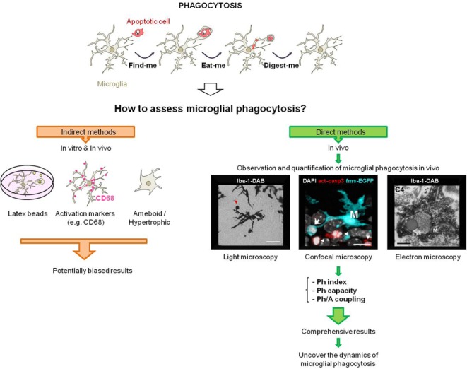
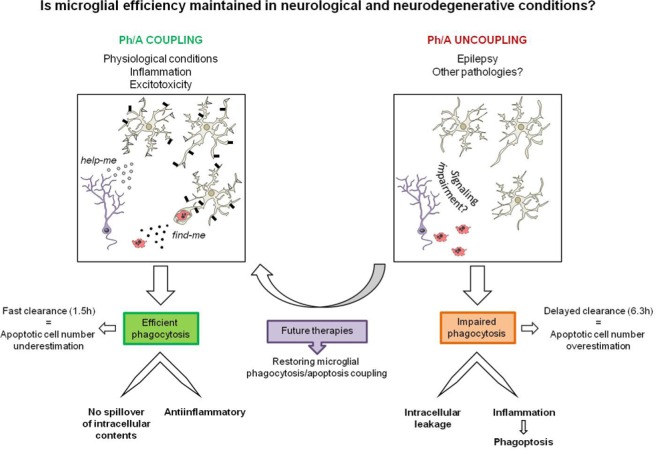
Apoptosis is a widespread phenomenon that occurs in the brain in both physiological and pathological conditions. Dead cells must be quickly removed to avoid the further toxic effects they exert in the parenchyma, a process executed by microglia, the brain professional phagocytes. Although phagocytosis is critical to maintain tissue homeostasis, it has long been either overlooked or indirectly assessed based on microglial morphology, expression of classical activation markers, or engulfment of artificial phagocytic targets . Nevertheless, these indirect methods present several limitations and, thus, direct observation and quantification of microglial phagocytosis is still necessary to fully grasp its relevance in the diseased brain. To overcome these caveats and obtain a comprehensive, quantitative picture of microglial phagocytosis we have developed a novel set of parameters. These parameters have allowed us to identify the different strategies utilized by microglia to cope with apoptotic challenges induced by excitotoxicity or inflammation. In contrast, we discovered that in mouse and human epilepsy microglia failed to find and engulf apoptotic cells, resulting in accumulation of debris and inflammation. Herein, we advocate that the efficiency of microglial phagocytosis should be routinely tested in neurodegenerative and neurological disorders, in order to determine the extent to which it contributes to apoptosis and inflammation found in these conditions. Finally, our findings point towards enhancing microglial phagocytosis as a novel therapeutic strategy to control tissue damage and inflammation, and accelerate recovery in brain diseases.
细胞凋亡是一种广泛存在的现象,在生理和病理条件下的大脑中都会发生。死亡细胞必须被迅速清除,以避免它们在实质组织中产生进一步的毒性作用,这一过程由小胶质细胞执行,小胶质细胞是大脑中的专业吞噬细胞。尽管吞噬作用对于维持组织稳态至关重要,但长期以来它要么被忽视,要么是基于小胶质细胞形态、经典激活标志物的表达或人工吞噬靶点的吞噬情况进行间接评估。然而,这些间接方法存在若干局限性,因此,要全面了解其在患病大脑中的相关性,仍有必要直接观察和量化小胶质细胞的吞噬作用。为了克服这些问题并获得小胶质细胞吞噬作用的全面定量情况,我们开发了一套新的参数。这些参数使我们能够识别小胶质细胞应对兴奋性毒性或炎症诱导的凋亡挑战所采用的不同策略。相比之下,我们发现,在小鼠和人类癫痫中,小胶质细胞无法找到并吞噬凋亡细胞,导致碎片堆积和炎症。在此,我们主张在神经退行性疾病和神经系统疾病中应常规检测小胶质细胞吞噬作用的效率,以确定其在这些疾病中对凋亡和炎症的影响程度。最后,我们的研究结果表明,增强小胶质细胞吞噬作用作为一种新的治疗策略,可控制组织损伤和炎症,并加速脑部疾病的恢复。