Department of Biochemistry, Radboud Institute for Molecular Life Sciences, Radboudumc, Nijmegen, The Netherlands; CIQUP/Department of Chemistry and Biochemistry, Faculty of Sciences, University of Porto, Porto, Portugal; Center for Neuroscience and Cell Biology (CNC), UC-Biotech, University of Coimbra, Coimbra, Portugal.
Department of Biochemistry, Radboud Institute for Molecular Life Sciences, Radboudumc, Nijmegen, The Netherlands.
Redox Biol. 2018 May;15:394-404. doi: 10.1016/j.redox.2017.12.018. Epub 2017 Dec 30.
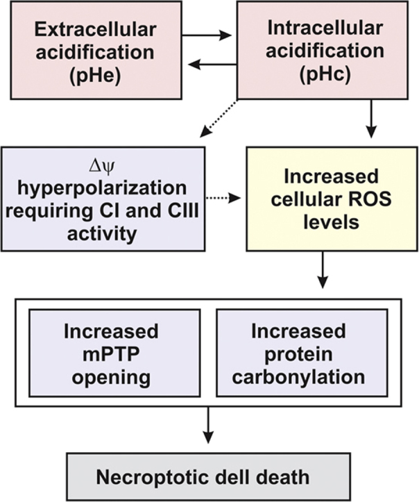
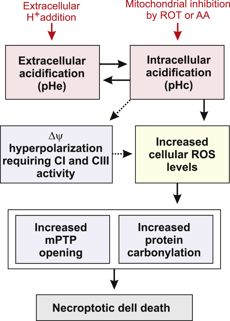
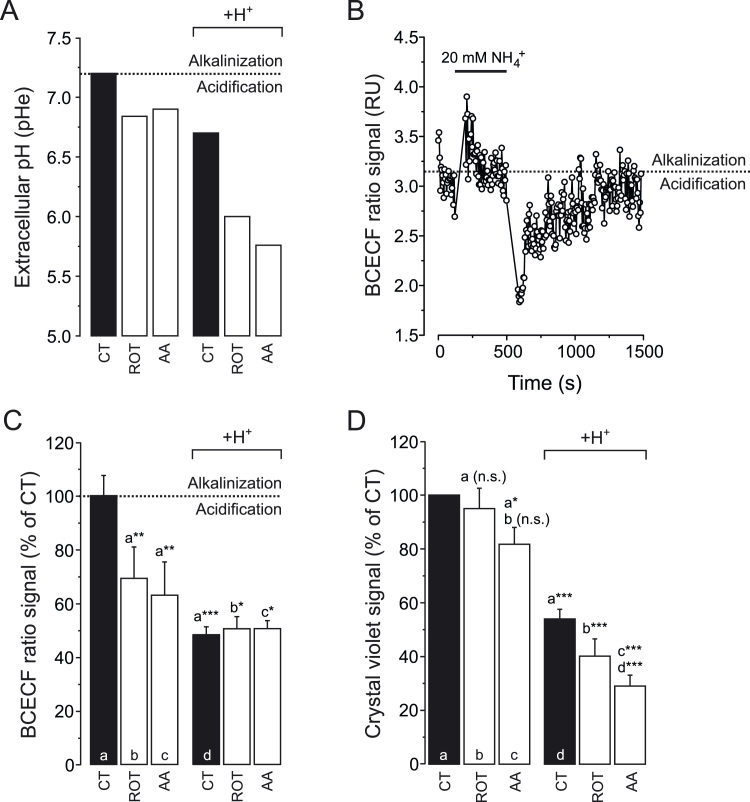
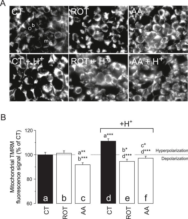
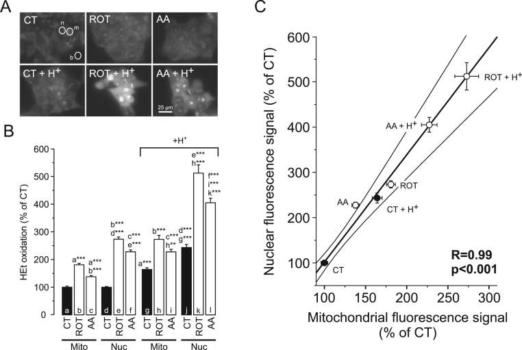
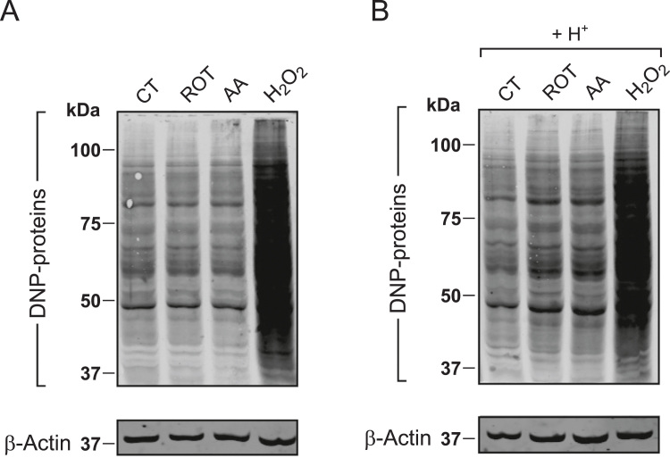
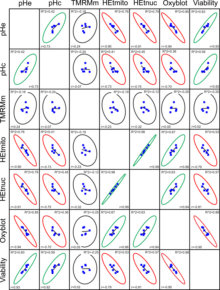
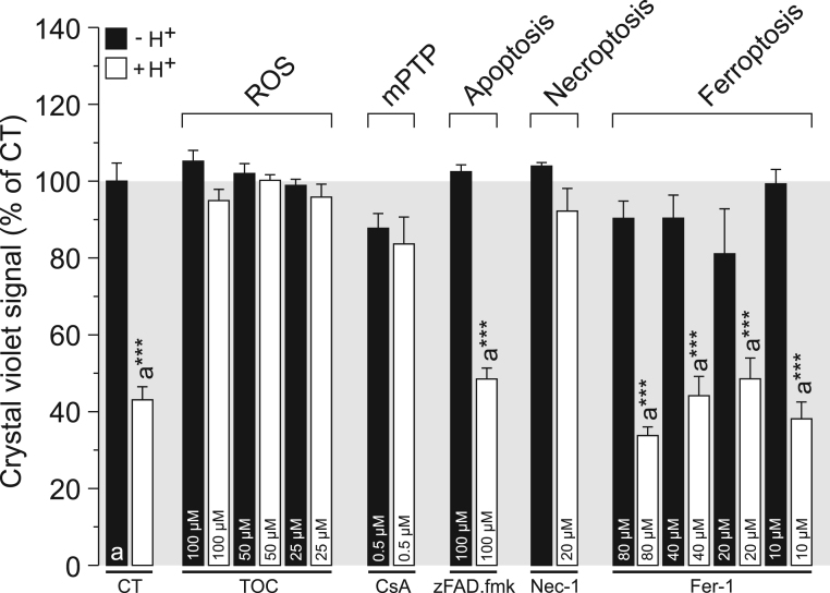
The extracellular pH (pHe) is a key determinant of the cellular (micro)environment and needs to be maintained within strict boundaries to allow normal cell function. Here we used HEK293 cells to study the effects of pHe acidification (24h), induced by mitochondrial inhibitors (rotenone, antimycin A) and/or extracellular HCl addition. Lowering pHe from 7.2 to 5.8 reduced cell viability by 70% and was paralleled by a decrease in cytosolic pH (pHc), hyperpolarization of the mitochondrial membrane potential (Δψ), increased levels of hydroethidine-oxidizing ROS and stimulation of protein carbonylation. Co-treatment with the antioxidant α-tocopherol, the mitochondrial permeability transition pore (mPTP) desensitizer cyclosporin A and Necrostatin-1, a combined inhibitor of Receptor-interacting serine/threonine-protein kinase 1 (RIPK1) and Indoleamine 2,3-dioxygenase (IDO), prevented acidification-induced cell death. In contrast, the caspase inhibitor zVAD.fmk and the ferroptosis inhibitor Ferrostatin-1 were ineffective. We conclude that extracellular acidification induces necroptotic cell death in HEK293 cells and that the latter involves intracellular acidification, mitochondrial functional impairment, increased ROS levels, mPTP opening and protein carbonylation. These findings suggest that acidosis of the extracellular environment (as observed in mitochondrial disorders, ischemia, acute inflammation and cancer) can induce cell death via a ROS- and mPTP opening-mediated pathogenic mechanism.
细胞外 pH 值(pHe)是细胞(微)环境的关键决定因素,需要在严格的范围内维持,以允许正常的细胞功能。在这里,我们使用 HEK293 细胞来研究由线粒体抑制剂(鱼藤酮、抗霉素 A)和/或细胞外 HCl 添加诱导的 pHe 酸化(24 小时)的影响。将 pHe 从 7.2 降低到 5.8 会使细胞活力降低 70%,同时细胞浆 pH 值(pHc)下降,线粒体膜电位(Δψ)去极化,羟乙基噻唑二唑(hydroethidine-oxidizing ROS)水平升高,蛋白质羰基化刺激。用抗氧化剂α-生育酚、线粒体通透性转换孔(mPTP)脱敏剂环孢菌素 A 和 Necrostatin-1(一种 Receptor-interacting serine/threonine-protein kinase 1(RIPK1)和吲哚胺 2,3-双加氧酶(IDO)的联合抑制剂)联合处理可预防酸化诱导的细胞死亡。相比之下,半胱氨酸天冬氨酸蛋白酶抑制剂 zVAD.fmk 和铁死亡抑制剂 Ferrostatin-1 无效。我们得出结论,细胞外酸化诱导 HEK293 细胞发生坏死性细胞死亡,后者涉及细胞内酸化、线粒体功能障碍、ROS 水平增加、mPTP 开放和蛋白质羰基化。这些发现表明,细胞外环境的酸中毒(如在线粒体疾病、缺血、急性炎症和癌症中观察到的)可以通过 ROS 和 mPTP 开放介导的致病机制诱导细胞死亡。