• 文献检索
  • 文档翻译
  • 深度研究
  • 学术资讯
  • Suppr Zotero 插件Zotero 插件
  • 邀请有礼
  • 套餐&价格
  • 历史记录
应用&插件
Suppr Zotero 插件Zotero 插件浏览器插件Mac 客户端Windows 客户端微信小程序
定价
高级版会员购买积分包购买API积分包
服务
文献检索文档翻译深度研究API 文档MCP 服务
关于我们
关于 Suppr公司介绍联系我们用户协议隐私条款
关注我们

Suppr 超能文献

核心技术专利:CN118964589B侵权必究
粤ICP备2023148730 号-1Suppr @ 2026

文献检索

告别复杂PubMed语法,用中文像聊天一样搜索,搜遍4000万医学文献。AI智能推荐,让科研检索更轻松。

立即免费搜索

文件翻译

保留排版,准确专业,支持PDF/Word/PPT等文件格式,支持 12+语言互译。

免费翻译文档

深度研究

AI帮你快速写综述,25分钟生成高质量综述,智能提取关键信息,辅助科研写作。

立即免费体验

连续块面扫描电子显微镜数据的分析与重建指南。

A guide to analysis and reconstruction of serial block face scanning electron microscopy data.

作者信息

Cocks E, Taggart M, Rind F C, White K

机构信息

Institute of Genetic Medicine, Newcastle University, Central Parkway, Newcastle upon Tyne, NE1 3BZ, UK.

Institute of Neuroscience, Newcastle University, Framlington Place, Newcastle upon Tyne, NE2 4HH, UK.

出版信息

J Microsc. 2018 May;270(2):217-234. doi: 10.1111/jmi.12676. Epub 2018 Jan 15.

DOI:10.1111/jmi.12676
PMID:29333754
原文链接:https://pmc.ncbi.nlm.nih.gov/articles/PMC5947172/
Abstract

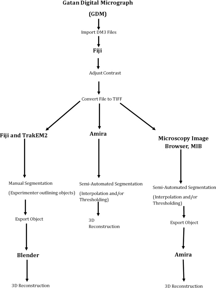
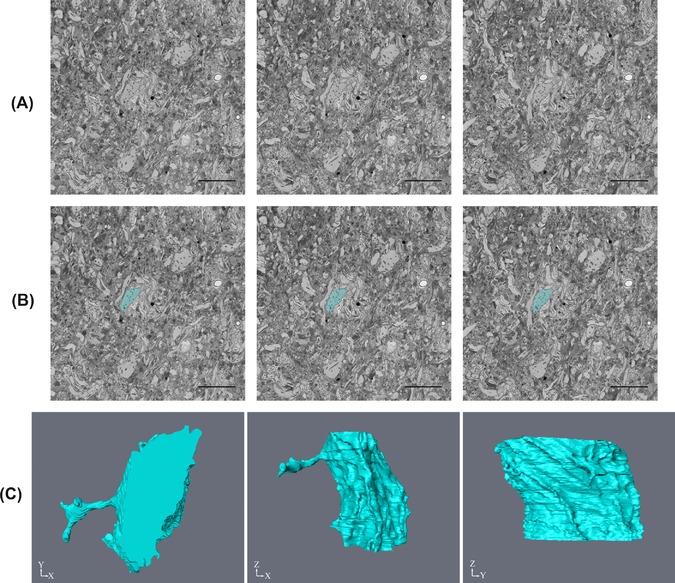
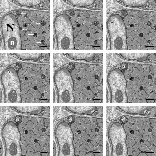
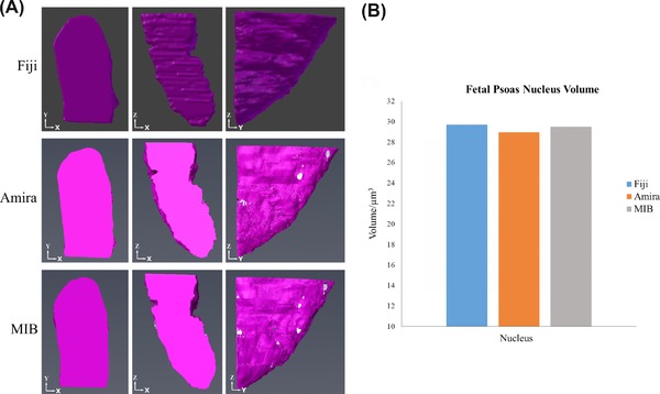
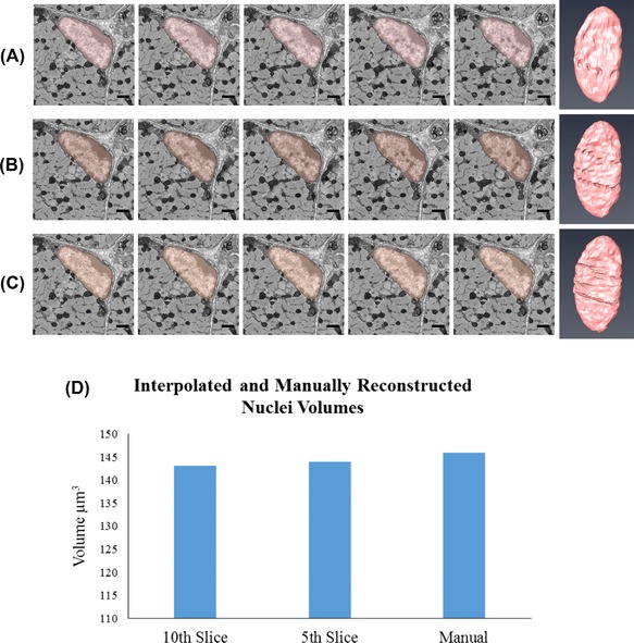
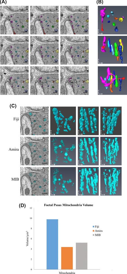
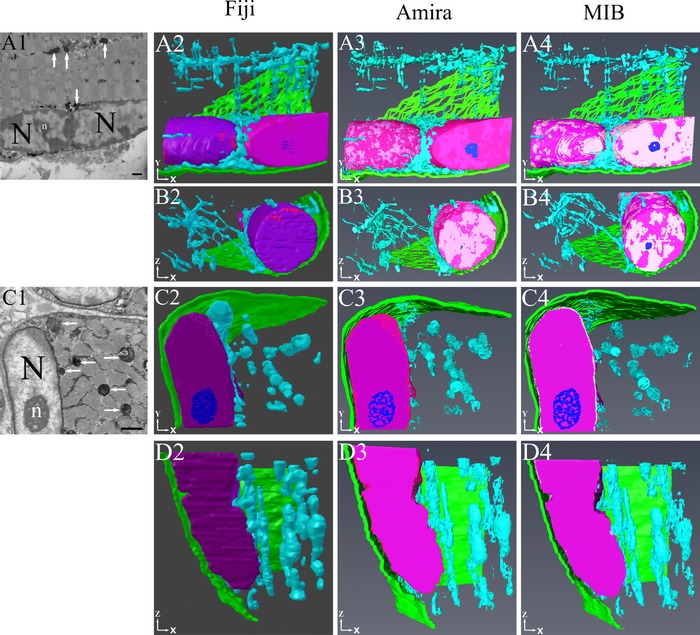
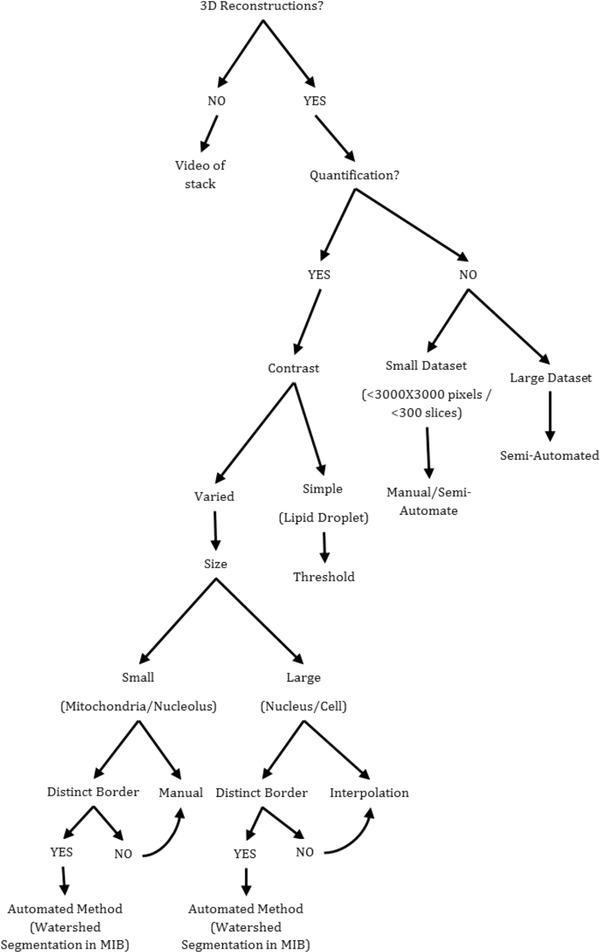
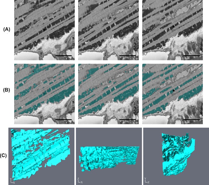
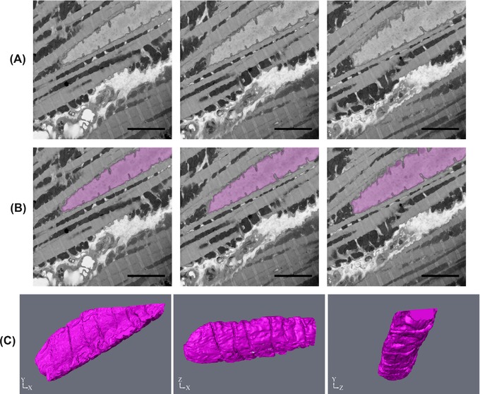
Serial block face scanning electron microscopy (SBF-SEM) is a relatively new technique that allows the acquisition of serially sectioned, imaged and digitally aligned ultrastructural data. There is a wealth of information that can be obtained from the resulting image stacks but this presents a new challenge for researchers - how to computationally analyse and make best use of the large datasets produced. One approach is to reconstruct structures and features of interest in 3D. However, the software programmes can appear overwhelming, time-consuming and not intuitive for those new to image analysis. There are a limited number of published articles that provide sufficient detail on how to do this type of reconstruction. Therefore, the aim of this paper is to provide a detailed step-by-step protocol, accompanied by tutorial videos, for several types of analysis programmes that can be used on raw SBF-SEM data, although there are more options available than can be covered here. To showcase the programmes, datasets of skeletal muscle from foetal and adult guinea pigs are initially used with procedures subsequently applied to guinea pig cardiac tissue and locust brain. The tissue is processed using the heavy metal protocol developed specifically for SBF-SEM. Trimmed resin blocks are placed into a Zeiss Sigma SEM incorporating the Gatan 3View and the resulting image stacks are analysed in three different programmes, Fiji, Amira and MIB, using a range of tools available for segmentation. The results from the image analysis comparison show that the analysis tools are often more suited to a particular type of structure. For example, larger structures, such as nuclei and cells, can be segmented using interpolation, which speeds up analysis; single contrast structures, such as the nucleolus, can be segmented using the contrast-based thresholding tools. Knowing the nature of the tissue and its specific structures (complexity, contrast, if there are distinct membranes, size) will help to determine the best method for reconstruction and thus maximize informative output from valuable tissue.

摘要

连续块面扫描电子显微镜(SBF-SEM)是一种相对较新的技术,它能够获取连续切片、成像并进行数字对齐的超微结构数据。从所得的图像堆栈中可以获得大量信息,但这给研究人员带来了新的挑战——如何通过计算分析并充分利用所产生的大型数据集。一种方法是对感兴趣的结构和特征进行三维重建。然而,对于图像分析新手来说,软件程序可能显得繁杂、耗时且不直观。关于如何进行此类重建的已发表文章数量有限。因此,本文的目的是提供一份详细的分步协议,并配有教程视频,介绍几种可用于原始SBF-SEM数据的分析程序,尽管可供选择的程序比本文所涵盖的要多。为了展示这些程序,最初使用了来自胎儿和成年豚鼠骨骼肌的数据集,随后将程序应用于豚鼠心脏组织和蝗虫大脑。使用专门为SBF-SEM开发的重金属协议对组织进行处理。将修整后的树脂块放入配备Gatan 3View的蔡司Sigma扫描电子显微镜中,然后使用一系列用于分割的工具,在三个不同的程序Fiji、Amira和MIB中对所得的图像堆栈进行分析。图像分析比较的结果表明,分析工具通常更适合特定类型的结构。例如,较大的结构,如细胞核和细胞,可以使用插值法进行分割,这可以加快分析速度;单一对比度的结构,如核仁,可以使用基于对比度的阈值工具进行分割。了解组织的性质及其特定结构(复杂性、对比度、是否有明显的膜、大小)将有助于确定最佳的重建方法,从而从宝贵的组织中最大化信息输出。

https://cdn.ncbi.nlm.nih.gov/pmc/blobs/6f2c/5947172/67cff0adc8c1/JMI-270-217-g012.jpg
https://cdn.ncbi.nlm.nih.gov/pmc/blobs/6f2c/5947172/f938061d1428/JMI-270-217-g001.jpg
https://cdn.ncbi.nlm.nih.gov/pmc/blobs/6f2c/5947172/0296e4531e3e/JMI-270-217-g002.jpg
https://cdn.ncbi.nlm.nih.gov/pmc/blobs/6f2c/5947172/344d0933dd1c/JMI-270-217-g003.jpg
https://cdn.ncbi.nlm.nih.gov/pmc/blobs/6f2c/5947172/9ddb72a55e7d/JMI-270-217-g004.jpg
https://cdn.ncbi.nlm.nih.gov/pmc/blobs/6f2c/5947172/36721dc428cf/JMI-270-217-g005.jpg
https://cdn.ncbi.nlm.nih.gov/pmc/blobs/6f2c/5947172/06330b0ab787/JMI-270-217-g006.jpg
https://cdn.ncbi.nlm.nih.gov/pmc/blobs/6f2c/5947172/89fc6d18066f/JMI-270-217-g007.jpg
https://cdn.ncbi.nlm.nih.gov/pmc/blobs/6f2c/5947172/ee539c5733f7/JMI-270-217-g008.jpg
https://cdn.ncbi.nlm.nih.gov/pmc/blobs/6f2c/5947172/9f94379722f7/JMI-270-217-g009.jpg
https://cdn.ncbi.nlm.nih.gov/pmc/blobs/6f2c/5947172/3f25195d472d/JMI-270-217-g010.jpg
https://cdn.ncbi.nlm.nih.gov/pmc/blobs/6f2c/5947172/97d05710b897/JMI-270-217-g011.jpg
https://cdn.ncbi.nlm.nih.gov/pmc/blobs/6f2c/5947172/67cff0adc8c1/JMI-270-217-g012.jpg

相似文献

1
A guide to analysis and reconstruction of serial block face scanning electron microscopy data.连续块面扫描电子显微镜数据的分析与重建指南。
J Microsc. 2018 May;270(2):217-234. doi: 10.1111/jmi.12676. Epub 2018 Jan 15.
2
A correlative approach for combining microCT, light and transmission electron microscopy in a single 3D scenario.一种将微计算机断层扫描、光学和透射电子显微镜在单个 3D 场景中组合的相关方法。
Front Zool. 2013 Aug 3;10(1):44. doi: 10.1186/1742-9994-10-44.
3
A workflow for 3D-CLEM investigating liver tissue.用于研究肝组织的 3D-CLEM 工作流程。
J Microsc. 2021 Mar;281(3):231-242. doi: 10.1111/jmi.12967. Epub 2020 Oct 27.
4
Serial block face scanning electron microscopy for the study of cardiac muscle ultrastructure at nanoscale resolutions.用于在纳米级分辨率下研究心肌超微结构的连续块面扫描电子显微镜技术。
J Mol Cell Cardiol. 2014 Nov;76:1-11. doi: 10.1016/j.yjmcc.2014.08.010. Epub 2014 Aug 20.
5
Protocols for Generating Surfaces and Measuring 3D Organelle Morphology Using Amira.使用 Amira 生成表面和测量 3D 细胞器形态的方案。
Cells. 2021 Dec 27;11(1):65. doi: 10.3390/cells11010065.
6
Investigation of heterocellular features of the mouse retinal neurovascular unit by 3D electron microscopy.利用 3D 电子显微镜研究小鼠视网膜神经血管单元的异细胞特征。
J Anat. 2023 Aug;243(2):245-257. doi: 10.1111/joa.13721. Epub 2022 Jul 16.
7
Development of protocols for the first serial block-face scanning electron microscopy (SBF SEM) studies of bone tissue.制定用于骨组织的第一代连续块面扫描电子显微镜 (SBF SEM) 研究的方案。
Bone. 2020 Feb;131:115107. doi: 10.1016/j.bone.2019.115107. Epub 2019 Oct 24.
8
Mitochondria morphometry in 3D datasets obtained from mouse brains with serial block-face scanning electron microscopy.使用连续块面扫描电子显微镜获取的小鼠脑 3D 数据集的线粒体形态计量学。
Methods Cell Biol. 2023;177:197-211. doi: 10.1016/bs.mcb.2023.01.021. Epub 2023 Mar 21.
9
Serial Block-Face Scanning Electron Microscopy (SBF-SEM) of Biological Tissue Samples.生物组织样品的连续块面扫描电子显微镜(SBF-SEM)观察。
J Vis Exp. 2021 Mar 26(169). doi: 10.3791/62045.
10
Semi-automatic stitching of filamentous structures in image stacks from serial-section electron tomography.序列切片电子断层扫描图像堆栈中丝状结构的半自动拼接。
J Microsc. 2021 Oct;284(1):25-44. doi: 10.1111/jmi.13039. Epub 2021 Jul 9.

引用本文的文献

1
Advances in Imaging Techniques for Mammalian/Human Ciliated Cell's Cilia: Insights into Structure, Function, and Dynamics.哺乳动物/人类纤毛细胞纤毛成像技术的进展:对结构、功能和动力学的见解
Biology (Basel). 2025 May 8;14(5):521. doi: 10.3390/biology14050521.
2
Layer-specific anatomical and physiological features of the retina's neurovascular unit.视网膜神经血管单元的层特异性解剖学和生理学特征。
Curr Biol. 2025 Jan 6;35(1):109-120.e4. doi: 10.1016/j.cub.2024.11.023. Epub 2024 Dec 16.
3
Unusual morphology of foveal Müller glia in an adult human born pre-term.

本文引用的文献

1
A Predictive 3D Multi-Scale Model of Biliary Fluid Dynamics in the Liver Lobule.肝脏小叶胆汁流体动力学的预测性三维多尺度模型。
Cell Syst. 2017 Mar 22;4(3):277-290.e9. doi: 10.1016/j.cels.2017.02.008. Epub 2017 Mar 18.
2
Volume scanning electron microscopy for imaging biological ultrastructure.用于生物超微结构成像的体积扫描电子显微镜。
Biol Cell. 2016 Nov;108(11):307-323. doi: 10.1111/boc.201600024. Epub 2016 Aug 12.
3
Serial block face scanning electron microscopy and the reconstruction of plant cell membrane systems.连续块面扫描电子显微镜术与植物细胞膜系统的重建
一名早产成年人类中黄斑区米勒胶质细胞的异常形态。
Front Cell Neurosci. 2024 Jun 27;18:1409405. doi: 10.3389/fncel.2024.1409405. eCollection 2024.
4
Unraveling the ultrastructure and dynamics of autophagic vesicles: Insights from advanced imaging techniques.解析自噬小泡的超微结构与动力学:先进成像技术带来的见解
FASEB Bioadv. 2024 May 2;6(7):189-199. doi: 10.1096/fba.2024-00035. eCollection 2024 Jul.
5
Correlative Imaging to Detect Rare HIV Reservoirs and Associated Damage in Tissues.相关性成像技术用于检测组织中罕见的 HIV 储存库及相关损伤。
Methods Mol Biol. 2024;2807:93-110. doi: 10.1007/978-1-0716-3862-0_7.
6
Volumetric Reconstruction of a Human Retinal Pigment Epithelial Cell Reveals Specialized Membranes and Polarized Distribution of Organelles.人视网膜色素上皮细胞的容积重建揭示了特化的膜和细胞器的极化分布。
Invest Ophthalmol Vis Sci. 2023 Dec 1;64(15):35. doi: 10.1167/iovs.64.15.35.
7
Unusual nuclear structures in male meiocytes of wild-type rye as revealed by volume microscopy.通过体视学方法显示野生型黑麦雄性减数分裂细胞中的异常核结构。
Ann Bot. 2023 Dec 5;132(6):1159-1174. doi: 10.1093/aob/mcad107.
8
Serial Block Face-Scanning Electron Microscopy as a Burgeoning Technology.序列块面扫描电子显微镜技术的兴起。
Adv Biol (Weinh). 2023 Aug;7(8):e2300139. doi: 10.1002/adbi.202300139. Epub 2023 May 28.
9
Investigation of heterocellular features of the mouse retinal neurovascular unit by 3D electron microscopy.利用 3D 电子显微镜研究小鼠视网膜神经血管单元的异细胞特征。
J Anat. 2023 Aug;243(2):245-257. doi: 10.1111/joa.13721. Epub 2022 Jul 16.
10
Three-dimensional imaging and analysis of the internal structure of SAPO-34 zeolite crystals.SAPO-34沸石晶体内部结构的三维成像与分析
RSC Adv. 2018 Oct 1;8(59):33631-33636. doi: 10.1039/c8ra05918g. eCollection 2018 Sep 28.
J Microsc. 2016 Aug;263(2):200-11. doi: 10.1111/jmi.12424. Epub 2016 May 20.
4
Anatomy and function of an excitatory network in the visual cortex.视觉皮层中一个兴奋性网络的解剖结构与功能
Nature. 2016 Apr 21;532(7599):370-4. doi: 10.1038/nature17192. Epub 2016 Mar 28.
5
Optimizing the 3D-reconstruction technique for serial block-face scanning electron microscopy.优化用于连续块面扫描电子显微镜的三维重建技术。
J Neurosci Methods. 2016 May 1;264:16-24. doi: 10.1016/j.jneumeth.2016.02.019. Epub 2016 Feb 27.
6
Reporting methods for processing and analysis of data from serial block face scanning electron microscopy.连续块面扫描电子显微镜数据处理与分析的报告方法
J Microsc. 2016 Jul;263(1):3-9. doi: 10.1111/jmi.12377. Epub 2016 Jan 21.
7
Microscopy Image Browser: A Platform for Segmentation and Analysis of Multidimensional Datasets.显微镜图像浏览器:一个用于多维数据集分割与分析的平台。
PLoS Biol. 2016 Jan 4;14(1):e1002340. doi: 10.1371/journal.pbio.1002340. eCollection 2016 Jan.
8
Saturated Reconstruction of a Volume of Neocortex.重建新皮层的体积
Cell. 2015 Jul 30;162(3):648-61. doi: 10.1016/j.cell.2015.06.054.
9
Connectome of the fly visual circuitry.果蝇视觉神经回路的连接组
Microscopy (Oxf). 2015 Feb;64(1):37-44. doi: 10.1093/jmicro/dfu102. Epub 2014 Dec 17.
10
NeuroMorph: a toolset for the morphometric analysis and visualization of 3D models derived from electron microscopy image stacks.NeuroMorph:一种用于对源自电子显微镜图像堆栈的3D模型进行形态计量分析和可视化的工具集。
Neuroinformatics. 2015 Jan;13(1):83-92. doi: 10.1007/s12021-014-9242-5.