VetImaging, VetCore Facility for Research, University of Veterinary Medicine, Veterinärplatz 1, 1210, Vienna, Austria.
Front Zool. 2013 Aug 3;10(1):44. doi: 10.1186/1742-9994-10-44.
In biomedical research, a huge variety of different techniques is currently available for the structural examination of small specimens, including conventional light microscopy (LM), transmission electron microscopy (TEM), confocal laser scanning microscopy (CLSM), microscopic X-ray computed tomography (microCT), and many others. Since every imaging method is physically limited by certain parameters, a correlative use of complementary methods often yields a significant broader range of information. Here we demonstrate the advantages of the correlative use of microCT, light microscopy, and transmission electron microscopy for the analysis of small biological samples.
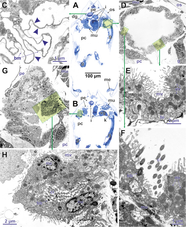
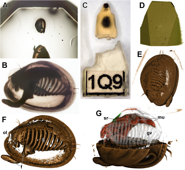
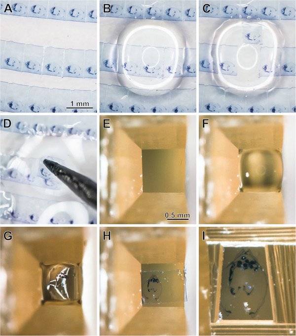
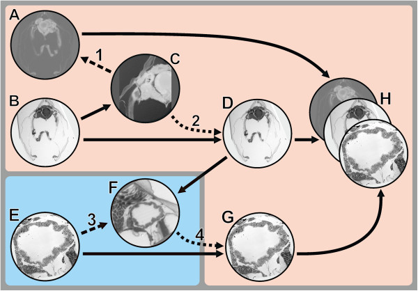
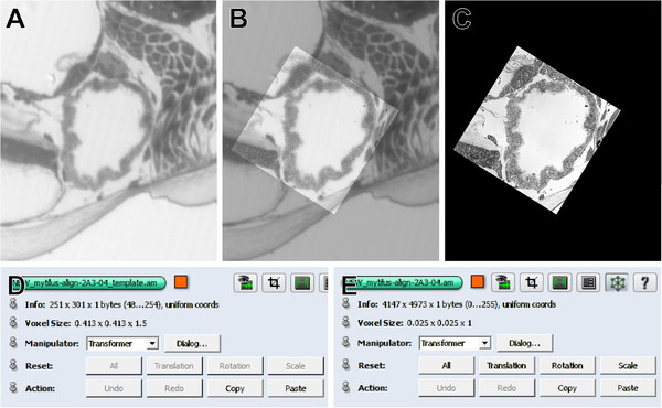
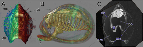
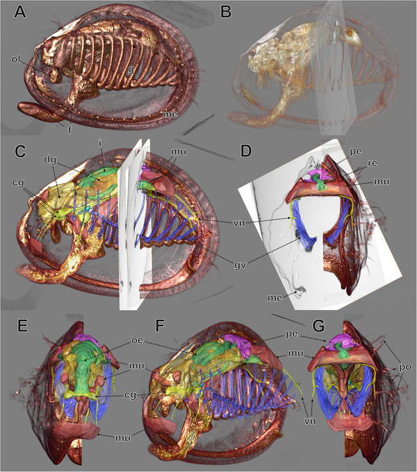
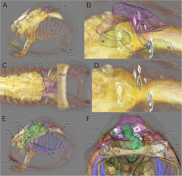
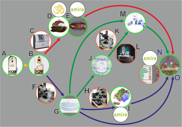
We used a small juvenile bivalve mollusc (Mytilus galloprovincialis, approximately 0.8 mm length) to demonstrate the workflow of a correlative examination by microCT, LM serial section analysis, and TEM-re-sectioning. Initially these three datasets were analyzed separately, and subsequently they were fused in one 3D scene. This workflow is very straightforward. The specimen was processed as usual for transmission electron microscopy including post-fixation in osmium tetroxide and embedding in epoxy resin. Subsequently it was imaged with microCT. Post-fixation in osmium tetroxide yielded sufficient X-ray contrast for microCT imaging, since the X-ray absorption of epoxy resin is low. Thereafter, the same specimen was serially sectioned for LM investigation. The serial section images were aligned and specific organ systems were reconstructed based on manual segmentation and surface rendering. According to the region of interest (ROI), specific LM sections were detached from the slides, re-mounted on resin blocks and re-sectioned (ultrathin) for TEM. For analysis, image data from the three different modalities was co-registered into a single 3D scene using the software AMIRA®. We were able to register both the LM section series volume and TEM slices neatly to the microCT dataset, with small geometric deviations occurring only in the peripheral areas of the specimen. Based on co-registered datasets the excretory organs, which were chosen as ROI for this study, could be investigated regarding both their ultrastructure as well as their position in the organism and their spatial relationship to adjacent tissues. We found structures typical for mollusc excretory systems, including ultrafiltration sites at the pericardial wall, and ducts leading from the pericardium towards the kidneys, which exhibit a typical basal infolding system.
The presented approach allows a comprehensive analysis and presentation of small objects regarding both the overall organization as well as cellular and subcellular details. Although our protocol involves a variety of different equipment and procedures, we maintain that it offers savings in both effort and cost. Co-registration of datasets from different imaging modalities can be accomplished with high-end desktop computers and offers new opportunities for understanding and communicating structural relationships within organisms and tissues. In general, the correlative use of different microscopic imaging techniques will continue to become more widespread in morphological and structural research in zoology. Classical TEM serial section investigations are extremely time consuming, and modern methods for 3D analysis of ultrastructure such as SBF-SEM and FIB-SEM are limited to very small volumes for examination. Thus the re-sectioning of LM sections is suitable for speeding up TEM examination substantially, while microCT could become a key-method for complementing ultrastructural examinations.
在生物医学研究中,目前有许多不同的技术可用于小标本的结构检查,包括常规的光学显微镜(LM)、透射电子显微镜(TEM)、共聚焦激光扫描显微镜(CLSM)、显微 X 射线计算机断层扫描(microCT)等。由于每种成像方法都受到某些参数的物理限制,因此互补方法的相关性使用通常会产生更广泛的信息。在这里,我们展示了 microCT、光显微镜和透射电子显微镜在分析小生物样本方面的相关性使用的优势。
我们使用了一只幼年双壳类软体动物(贻贝,长约 0.8 毫米)来演示 microCT、LM 连续切片分析和 TEM 重切的相关性检查工作流程。最初,这三个数据集分别进行了分析,然后将它们融合在一个 3D 场景中。该工作流程非常简单。标本按照透射电子显微镜的常规方法进行处理,包括锇四氧化后固定和环氧树脂包埋。随后,对其进行 microCT 成像。锇四氧化后的后固定为 microCT 成像提供了足够的 X 射线对比度,因为环氧树脂的 X 射线吸收率很低。此后,对同一样本进行连续切片以进行 LM 研究。对连续切片图像进行对齐,并根据手动分割和表面渲染对特定器官系统进行重建。根据感兴趣区域(ROI),从幻灯片上取下特定的 LM 切片,重新安装在树脂块上并重新切片(超薄)进行 TEM。对于分析,使用 AMIRA®软件将来自三种不同模式的图像数据配准到单个 3D 场景中。我们能够将 LM 切片系列体积和 TEM 切片整齐地配准到 microCT 数据集,只有在标本的外围区域才会出现小的几何偏差。基于配准数据集,可以研究选择作为该研究的 ROI 的排泄器官,同时研究其超微结构以及在生物体中的位置及其与相邻组织的空间关系。我们发现了一些典型的软体动物排泄系统结构,包括心壁上的超滤位点,以及从心脏通向肾脏的导管,这些结构表现出典型的基底内折系统。
该方法允许对小物体进行全面的分析和呈现,包括整体组织以及细胞和亚细胞细节。尽管我们的方案涉及多种不同的设备和程序,但我们认为它在节省时间和成本方面具有优势。来自不同成像模式的数据集的配准可以使用高端台式计算机来完成,并为理解和交流生物体和组织内的结构关系提供新的机会。一般来说,不同显微镜成像技术的相关性使用将继续在动物学形态学和结构研究中得到更广泛的应用。经典的 TEM 连续切片研究非常耗时,而用于超微结构 3D 分析的现代方法,如 SBF-SEM 和 FIB-SEM,由于体积限制,只能检查非常小的体积。因此,LM 切片的重新切片非常适合大大加快 TEM 检查速度,而 microCT 则可以成为补充超微结构检查的关键方法。