Díaz-Valdivia Natalia I, Calderón Claudia C, Díaz Jorge E, Lobos-González Lorena, Sepulveda Hugo, Ortíz Rina J, Martinez Samuel, Silva Veronica, Maldonado Horacio J, Silva Patricio, Wehinger Sergio, Burzio Verónica A, Torres Vicente A, Montecino Martín, Leyton Lisette, Quest Andrew F G
Cellular Communication Laboratory, Center for Molecular Studies of the Cell (CEMC), Advanced Center for Chronic Diseases (ACCDiS), Faculty of Medicine, Universidad de Chile, Santiago, Chile.
Institute for Research in Dental Sciences, Faculty of Dentistry, Universidad de Chile, Santiago, Chile.
Oncotarget. 2017 Dec 5;8(67):111943-111965. doi: 10.18632/oncotarget.22955. eCollection 2017 Dec 19.
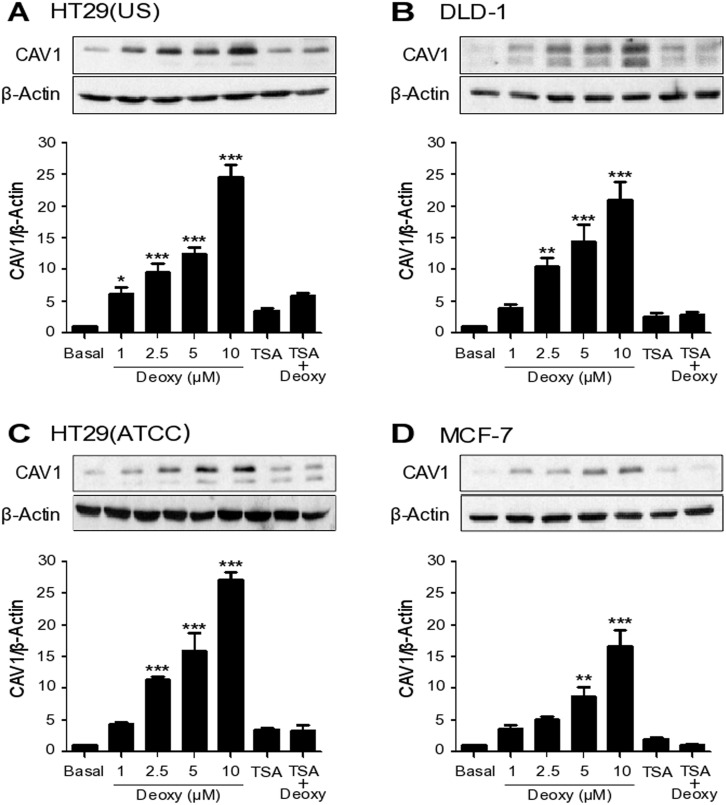
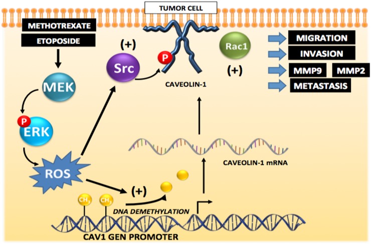
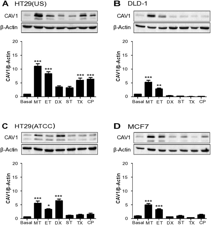
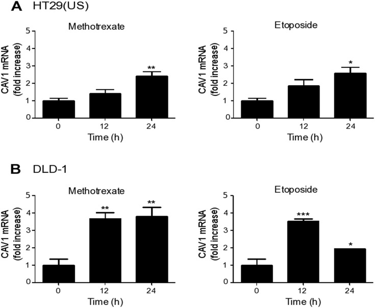
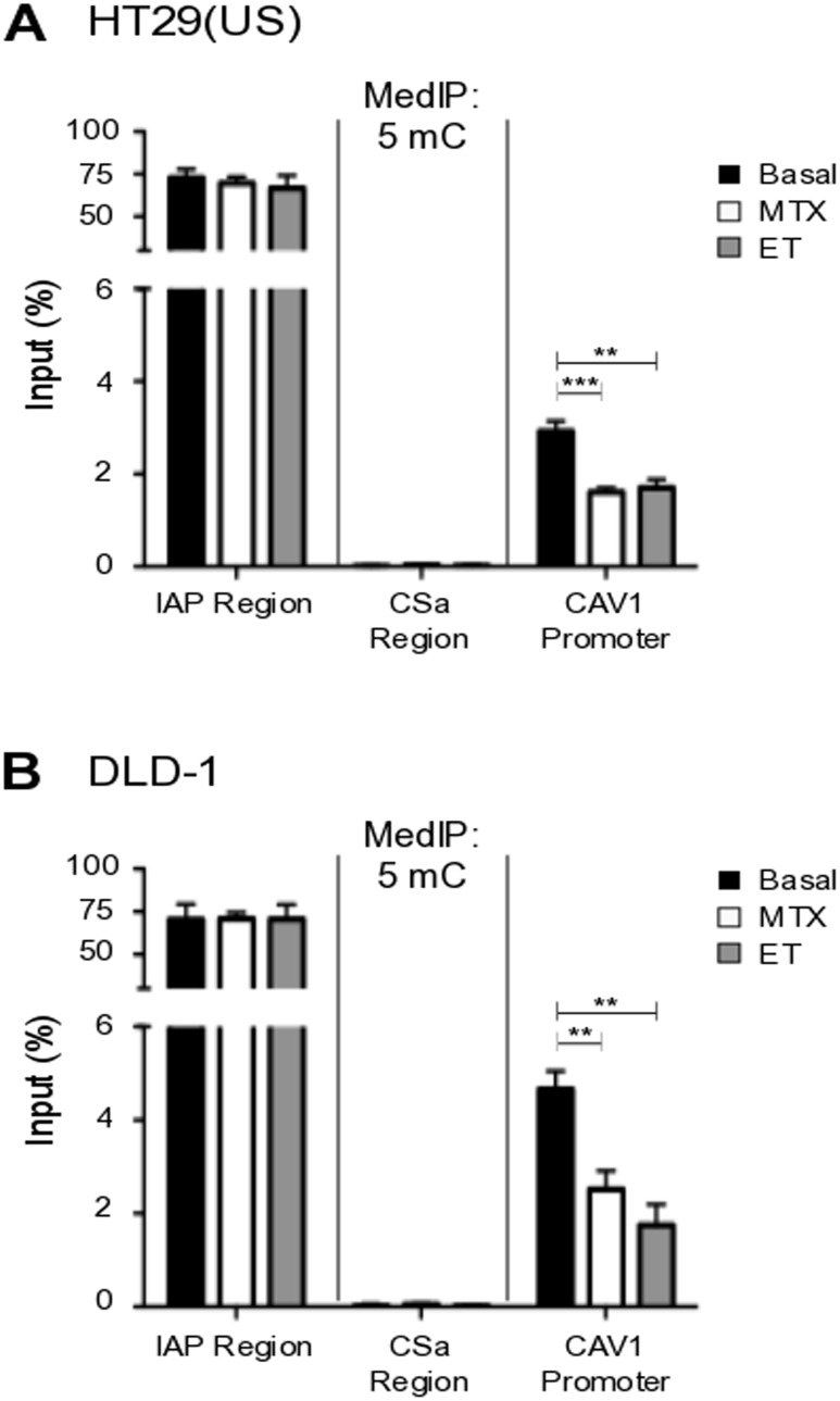
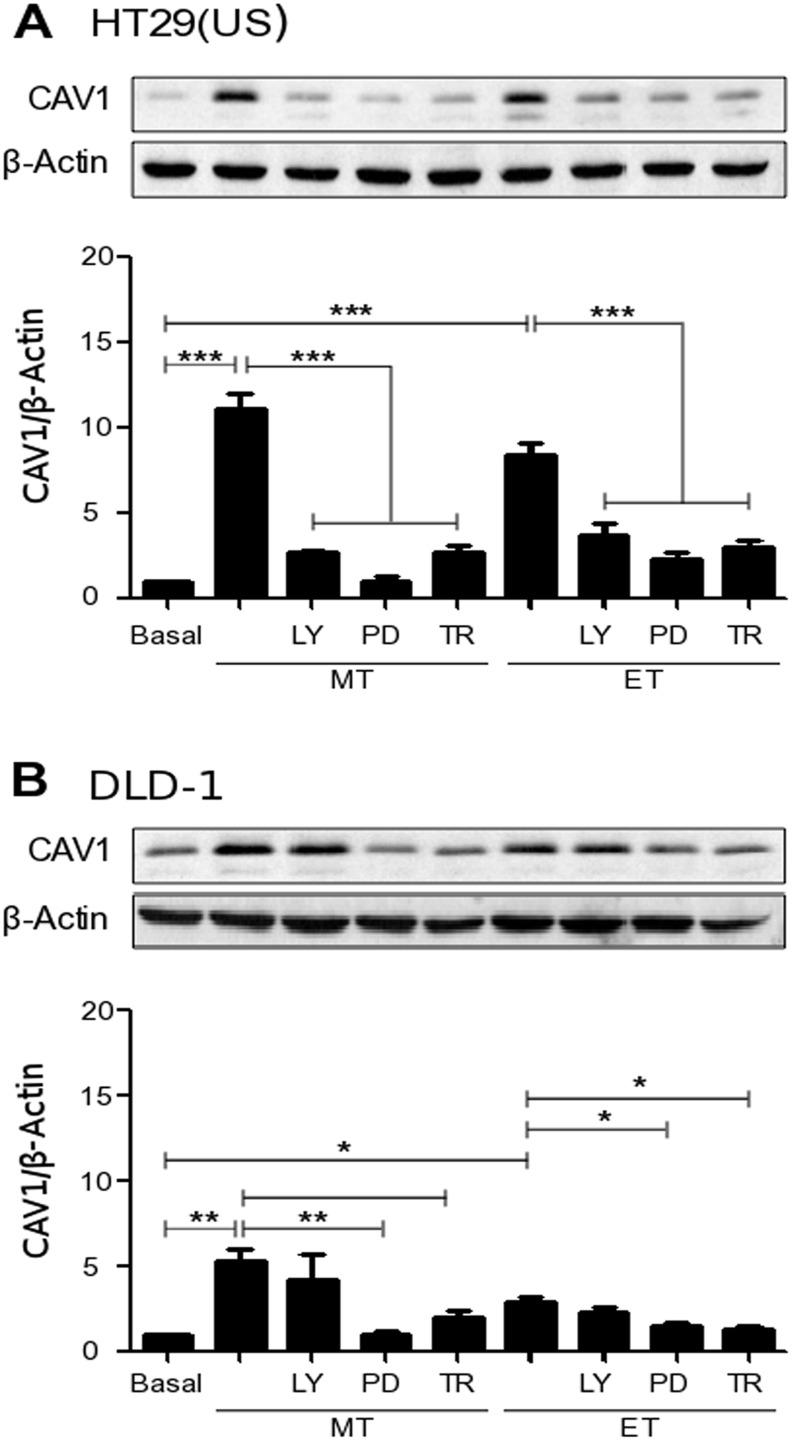
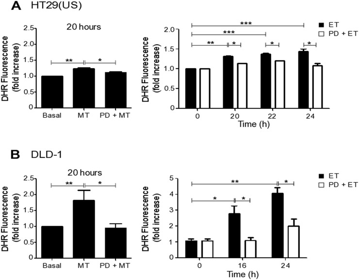
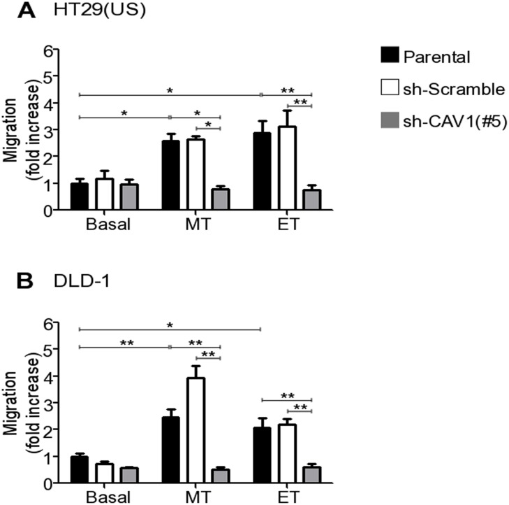
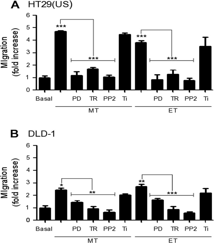
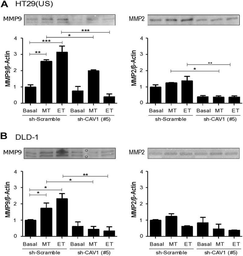
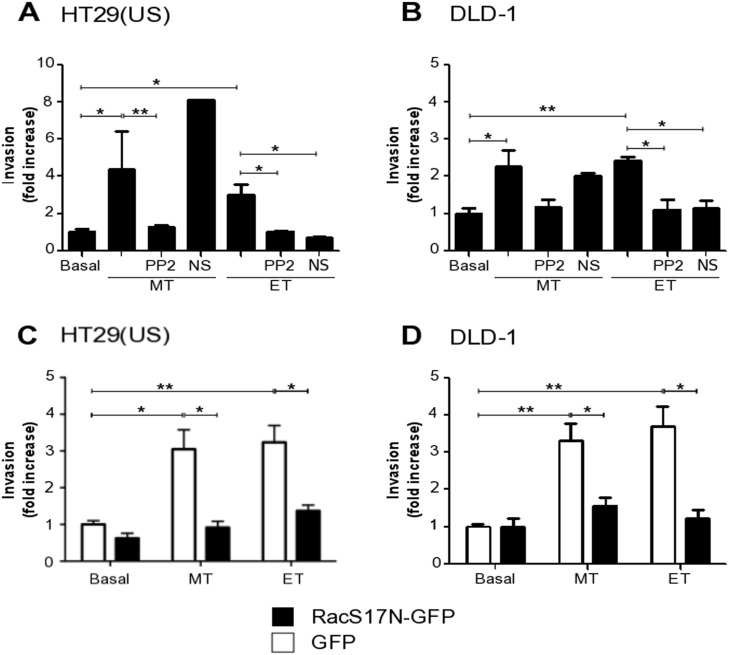
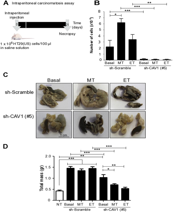
Expression of the scaffolding protein Caveolin-1 (CAV1) enhances migration and invasion of metastatic cancer cells. Yet, CAV1 also functions as a tumor suppressor in early stages of cancer, where expression is suppressed by epigenetic mechanisms. Thus, we sought to identify stimuli/mechanisms that revert epigenetic CAV1 silencing in cancer cells and evaluate how this affects their metastatic potential. We reasoned that restricted tissue availability of anti-neoplastic drugs during chemotherapy might expose cancer cells to sub-therapeutic concentrations, which activate signaling pathways and the expression of CAV1 to favor the acquisition of more aggressive traits. Here, we used [2D, invasion] and (metastasis) assays, as well as genetic and biochemical approaches to address this question. Colon and breast cancer cells were identified where CAV1 levels were low due to epigenetic suppression and could be reverted by treatment with the methyltransferase inhibitor 5'-azacytidine. Exposure of these cells to anti-neoplastic drugs for short periods of time (24-48 h) increased CAV1 expression through ROS production and MEK/ERK activation. In colon cancer cells, increased CAV1 expression enhanced migration and invasion via pathways requiring Src-family kinases, as well as Rac-1 activity. Finally, elevated CAV1 expression in colon cancer cells following exposure to sub-cytotoxic drug concentrations increased their metastatic potential . Therefore exposure of cancer cells to anti-neoplastic drugs at non-lethal drug concentrations induces signaling events and changes in transcription that favor CAV1-dependent migration, invasion and metastasis. Importantly, this may occur in the absence of selection for drug-resistance.
支架蛋白小窝蛋白-1(CAV1)的表达增强转移性癌细胞的迁移和侵袭能力。然而,CAV1在癌症早期也发挥肿瘤抑制作用,其表达在此阶段通过表观遗传机制受到抑制。因此,我们试图确定能逆转癌细胞中CAV1表观遗传沉默的刺激因素/机制,并评估这对其转移潜能有何影响。我们推断,化疗期间抗肿瘤药物的组织可利用性受限可能使癌细胞暴露于亚治疗浓度,从而激活信号通路和CAV1的表达,促使癌细胞获得更具侵袭性的特征。在此,我们使用二维侵袭试验和转移试验,以及遗传学和生物化学方法来解决这个问题。我们鉴定出由于表观遗传抑制导致CAV1水平较低且可通过甲基转移酶抑制剂5'-氮杂胞苷处理而逆转的结肠癌细胞和乳腺癌细胞。这些细胞短时间(24 - 48小时)暴露于抗肿瘤药物通过活性氧生成和MEK/ERK激活增加了CAV1的表达。在结肠癌细胞中,增加的CAV1表达通过需要Src家族激酶以及Rac - 1活性的途径增强了迁移和侵袭能力。最后,结肠癌细胞在暴露于亚细胞毒性药物浓度后CAV1表达升高增加了它们的转移潜能。因此,癌细胞在非致死药物浓度下暴露于抗肿瘤药物会诱导信号事件和转录变化,有利于依赖CAV1的迁移、侵袭和转移。重要的是,这可能在没有选择耐药性的情况下发生。