• 文献检索
  • 文档翻译
  • 深度研究
  • 学术资讯
  • Suppr Zotero 插件Zotero 插件
  • 邀请有礼
  • 套餐&价格
  • 历史记录
应用&插件
Suppr Zotero 插件Zotero 插件浏览器插件Mac 客户端Windows 客户端微信小程序
定价
高级版会员购买积分包购买API积分包
服务
文献检索文档翻译深度研究API 文档MCP 服务
关于我们
关于 Suppr公司介绍联系我们用户协议隐私条款
关注我们

Suppr 超能文献

核心技术专利:CN118964589B侵权必究
粤ICP备2023148730 号-1Suppr @ 2026

文献检索

告别复杂PubMed语法,用中文像聊天一样搜索,搜遍4000万医学文献。AI智能推荐,让科研检索更轻松。

立即免费搜索

文件翻译

保留排版,准确专业,支持PDF/Word/PPT等文件格式,支持 12+语言互译。

免费翻译文档

深度研究

AI帮你快速写综述,25分钟生成高质量综述,智能提取关键信息,辅助科研写作。

立即免费体验

视神经挤压伤后再生重塑损伤部位:成像质谱技术的分子见解。

Optic Nerve Regeneration After Crush Remodels the Injury Site: Molecular Insights From Imaging Mass Spectrometry.

机构信息

Stein Eye Institute, David Geffen School of Medicine at UCLA, Los Angeles, California, United States.

Vanderbilt Mass Spectrometry Research Center and Department of Biochemistry, Vanderbilt University School of Medicine, Nashville, Tennessee, United States.

出版信息

Invest Ophthalmol Vis Sci. 2018 Jan 1;59(1):212-222. doi: 10.1167/iovs.17-22509.

DOI:10.1167/iovs.17-22509
PMID:29340649
原文链接:https://pmc.ncbi.nlm.nih.gov/articles/PMC5770179/
Abstract

PURPOSE

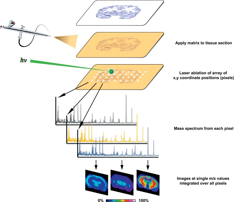
Mammalian central nervous system axons fail to regenerate after injury. Contributing factors include limited intrinsic growth capacity and an inhibitory glial environment. Inflammation-induced optic nerve regeneration (IIR) is thought to boost retinal ganglion cell (RGC) intrinsic growth capacity through progrowth gene expression, but effects on the inhibitory glial environment of the optic nerve are unexplored. To investigate progrowth molecular changes associated with reactive gliosis during IIR, we developed an imaging mass spectrometry (IMS)-based approach that identifies discriminant molecular signals in and around optic nerve crush (ONC) sites.

METHODS

ONC was performed in rats, and IIR was established by intravitreal injection of a yeast cell wall preparation. Optic nerves were collected at various postcrush intervals, and longitudinal sections were analyzed with matrix-assisted laser desorption/ionization (MALDI) IMS and data mining. Immunohistochemistry and confocal microscopy were used to compare discriminant molecular features with cellular features of reactive gliosis.

RESULTS

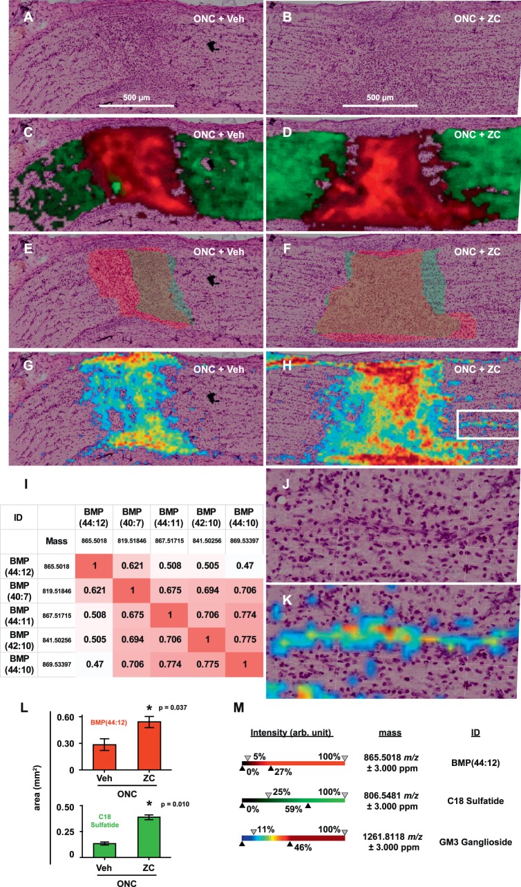
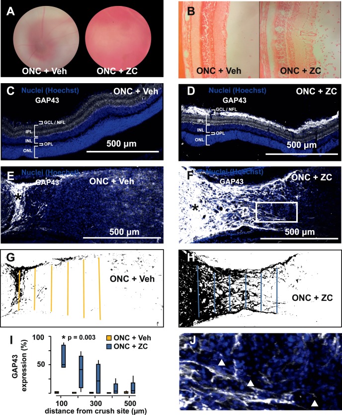
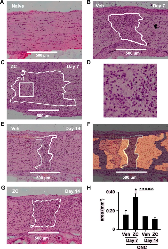
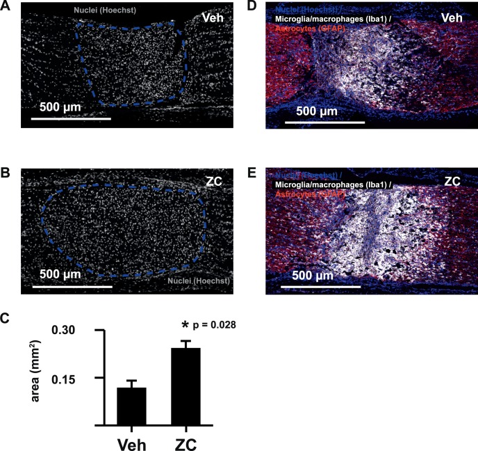
IIR increased the area of the crush site that was occupied by a dense cellular infiltrate and mass spectral features consistent with lysosome-specific lipids. IIR also increased immunohistochemical labeling for microglia and macrophages. IIR enhanced clearance of lipid sulfatide myelin-associated inhibitors of axon growth and accumulation of simple GM3 gangliosides in a spatial distribution consistent with degradation of plasma membrane from degenerated axons.

CONCLUSIONS

IIR promotes a robust phagocytic response that improves clearance of myelin and axon debris. This growth-permissive molecular remodeling of the crush injury site extends our current understanding of IIR to include mechanisms extrinsic to the RGC.

摘要

目的

哺乳动物中枢神经系统轴突在损伤后无法再生。其致病因素包括内在生长能力有限和抑制性神经胶质环境。炎症诱导的视神经再生(IIR)被认为通过生长基因表达来增强视网膜神经节细胞(RGC)的内在生长能力,但对视神经抑制性神经胶质环境的影响尚未得到探索。为了研究与 IIR 中反应性神经胶质相关的促生长分子变化,我们开发了一种基于成像质谱(IMS)的方法,该方法可识别视神经挤压(ONC)部位及其周围的有区别的分子信号。

方法

在大鼠中进行 ONC,通过眼内注射酵母细胞壁制剂来建立 IIR。在各种挤压后间隔收集视神经,并通过基质辅助激光解吸/电离(MALDI)IMS 和数据挖掘对纵向切片进行分析。免疫组织化学和共聚焦显微镜用于比较有区别的分子特征与反应性神经胶质的细胞特征。

结果

IIR 增加了挤压部位的面积,该部位被密集的细胞浸润和与溶酶体特异性脂质一致的质谱特征所占据。IIR 还增加了小胶质细胞和巨噬细胞的免疫组织化学标记。IIR 增强了脂质硫脂髓鞘轴突生长抑制剂的清除和简单 GM3 神经节苷脂的积累,其空间分布与从退化轴突降解的质膜一致。

结论

IIR 促进了强大的吞噬反应,改善了髓鞘和轴突碎片的清除。挤压损伤部位的这种生长促进的分子重塑扩展了我们对 IIR 的现有理解,包括 RGC 以外的机制。

https://cdn.ncbi.nlm.nih.gov/pmc/blobs/3779/5770179/c0ea578659fc/i1552-5783-59-1-212-f05.jpg
https://cdn.ncbi.nlm.nih.gov/pmc/blobs/3779/5770179/ecdec045f058/i1552-5783-59-1-212-f01.jpg
https://cdn.ncbi.nlm.nih.gov/pmc/blobs/3779/5770179/b6c28bd2878c/i1552-5783-59-1-212-f02.jpg
https://cdn.ncbi.nlm.nih.gov/pmc/blobs/3779/5770179/363883cedd3e/i1552-5783-59-1-212-f03.jpg
https://cdn.ncbi.nlm.nih.gov/pmc/blobs/3779/5770179/d31b11f3b530/i1552-5783-59-1-212-f04.jpg
https://cdn.ncbi.nlm.nih.gov/pmc/blobs/3779/5770179/c0ea578659fc/i1552-5783-59-1-212-f05.jpg

相似文献

1
Optic Nerve Regeneration After Crush Remodels the Injury Site: Molecular Insights From Imaging Mass Spectrometry.视神经挤压伤后再生重塑损伤部位:成像质谱技术的分子见解。
Invest Ophthalmol Vis Sci. 2018 Jan 1;59(1):212-222. doi: 10.1167/iovs.17-22509.
2
Regenerative Responses and Axon Pathfinding of Retinal Ganglion Cells in Chronically Injured Mice.慢性损伤小鼠视网膜神经节细胞的再生反应与轴突寻路
Invest Ophthalmol Vis Sci. 2017 Mar 1;58(3):1743-1750. doi: 10.1167/iovs.16-19873.
3
Topical administration of a Rock/Net inhibitor promotes retinal ganglion cell survival and axon regeneration after optic nerve injury.局部应用Rock/Net抑制剂可促进视神经损伤后视网膜神经节细胞存活和轴突再生。
Exp Eye Res. 2017 May;158:33-42. doi: 10.1016/j.exer.2016.07.006. Epub 2016 Jul 18.
4
Attenuation of Axonal Degeneration by Calcium Channel Inhibitors Improves Retinal Ganglion Cell Survival and Regeneration After Optic Nerve Crush.钙通道抑制剂减轻轴突退变可改善视神经挤压后视网膜神经节细胞的存活与再生。
Mol Neurobiol. 2017 Jan;54(1):72-86. doi: 10.1007/s12035-015-9676-2. Epub 2016 Jan 5.
5
Immunohistochemical localization of CNTFRalpha in adult mouse retina and optic nerve following intraorbital nerve crush: evidence for the axonal loss of a trophic factor receptor after injury.眶内神经挤压后成年小鼠视网膜和视神经中CNTFRalpha的免疫组织化学定位:损伤后营养因子受体轴突损失的证据。
J Comp Neurol. 2007 Jan 10;500(2):384-400. doi: 10.1002/cne.21174.
6
Distribution of mesenchymal stem cells and effects on neuronal survival and axon regeneration after optic nerve crush and cell therapy.视神经挤压伤及细胞治疗后间充质干细胞的分布及其对神经元存活和轴突再生的影响
PLoS One. 2014 Oct 27;9(10):e110722. doi: 10.1371/journal.pone.0110722. eCollection 2014.
7
Macrophage-derived factors stimulate optic nerve regeneration.巨噬细胞衍生因子刺激视神经再生。
J Neurosci. 2003 Mar 15;23(6):2284-93. doi: 10.1523/JNEUROSCI.23-06-02284.2003.
8
Activation of the BMP4/Smad1 Pathway Promotes Retinal Ganglion Cell Survival and Axon Regeneration.BMP4/Smad1 通路的激活促进视网膜神经节细胞存活和轴突再生。
Invest Ophthalmol Vis Sci. 2019 Apr 1;60(5):1748-1759. doi: 10.1167/iovs.18-26449.
9
Sema-3A indirectly disrupts the regeneration process of goldfish optic nerve after controlled injury.Sema-3A 可间接破坏控制损伤后金鱼视神经的再生过程。
Graefes Arch Clin Exp Ophthalmol. 2010 Oct;248(10):1423-35. doi: 10.1007/s00417-010-1377-y. Epub 2010 May 7.
10
A Novel Rat Model with Long Range Optic Nerve Injury to Study Retinal Ganglion Cells Endogenous Regeneration.一种新型长程视神经损伤大鼠模型用于研究视网膜神经节细胞内源性再生。
Neuroscience. 2021 Jun 15;465:71-84. doi: 10.1016/j.neuroscience.2021.04.014. Epub 2021 Apr 23.

引用本文的文献

1
Extracellular Vesicle Metabolomics Holds Promise for Adult Axon Regeneration.细胞外囊泡代谢组学有望促进成体轴突再生。
Metabolites. 2025 Jul 4;15(7):454. doi: 10.3390/metabo15070454.
2
Evaluating the Evidence for Neuroprotective and Axonal Regenerative Activities of Different Inflammatory Cell Types After Optic Nerve Injury.评估视神经损伤后不同炎症细胞类型的神经保护和轴突再生活性的证据。
Mol Neurobiol. 2025 May;62(5):6212-6227. doi: 10.1007/s12035-024-04679-3. Epub 2024 Dec 30.
3
Interleukin-4 protects retinal ganglion cells and promotes axon regeneration.

本文引用的文献

1
Imaging MS of Rodent Ocular Tissues and the Optic Nerve.啮齿动物眼部组织和视神经的成像质谱分析
Methods Mol Biol. 2017;1618:15-27. doi: 10.1007/978-1-4939-7051-3_3.
2
Reassembly of Excitable Domains after CNS Axon Regeneration.中枢神经系统轴突再生后兴奋性区域的重新组装。
J Neurosci. 2016 Aug 31;36(35):9148-60. doi: 10.1523/JNEUROSCI.1747-16.2016.
3
Neural activity promotes long-distance, target-specific regeneration of adult retinal axons.神经活动促进成年视网膜轴突的长距离、靶向特异性再生。
白细胞介素-4可保护视网膜神经节细胞并促进轴突再生。
Cell Commun Signal. 2024 Apr 22;22(1):236. doi: 10.1186/s12964-024-01604-y.
4
Lysolipids are prominent in subretinal drusenoid deposits, a high-risk phenotype in age-related macular degeneration.溶血磷脂在视网膜下玻璃膜疣状沉积物中很突出,这是年龄相关性黄斑变性的一种高风险表型。
Front Ophthalmol (Lausanne). 2023;3. doi: 10.3389/fopht.2023.1258734. Epub 2023 Nov 24.
5
High-Resolution Imaging Mass Spectrometry of Human Donor Eye: Photoreceptors Cells and Basal Laminar Deposit of Age-Related Macular Degeneration.高分辨率成像质谱分析人眼捐献组织:年龄相关性黄斑变性的感光细胞和基底膜层沉积物。
Adv Exp Med Biol. 2023;1415:3-7. doi: 10.1007/978-3-031-27681-1_1.
6
The role of monocytes in optic nerve injury.单核细胞在视神经损伤中的作用。
Neural Regen Res. 2023 Aug;18(8):1666-1671. doi: 10.4103/1673-5374.363825.
7
Intraocular Delivery of a Collagen Mimetic Peptide Repairs Retinal Ganglion Cell Axons in Chronic and Acute Injury Models.胶原模拟肽经眼内递送至慢性和急性损伤模型中修复视网膜神经节细胞轴突。
Int J Mol Sci. 2022 Mar 8;23(6):2911. doi: 10.3390/ijms23062911.
8
Traumatic optic neuropathy: a review of current studies.外伤性视神经病变:当前研究综述。
Neurosurg Rev. 2022 Jun;45(3):1895-1913. doi: 10.1007/s10143-021-01717-9. Epub 2022 Jan 16.
9
Developmentally upregulated transcriptional elongation factor a like 3 suppresses axon regeneration after optic nerve injury.发育上调的转录延伸因子 A 样 3 抑制视神经损伤后的轴突再生。
Neurosci Lett. 2021 Nov 20;765:136260. doi: 10.1016/j.neulet.2021.136260. Epub 2021 Sep 21.
10
Lipid Landscape of the Human Retina and Supporting Tissues Revealed by High-Resolution Imaging Mass Spectrometry.高分辨率成像质谱技术揭示的人视网膜及其支持组织的脂质全景图。
J Am Soc Mass Spectrom. 2020 Dec 2;31(12):2426-2436. doi: 10.1021/jasms.0c00119. Epub 2020 Jul 24.
Nat Neurosci. 2016 Aug;19(8):1073-84. doi: 10.1038/nn.4340. Epub 2016 Jul 11.
4
Sphingosine 1-phosphate receptor 1 is required for retinal ganglion cell survival after optic nerve trauma.1-磷酸鞘氨醇受体1是视神经损伤后视网膜神经节细胞存活所必需的。
J Neurochem. 2016 Aug;138(4):571-86. doi: 10.1111/jnc.13701. Epub 2016 Jul 6.
5
Intrinsic Control of Axon Regeneration.轴突再生的内在控制。
Neuron. 2016 May 4;90(3):437-51. doi: 10.1016/j.neuron.2016.04.022.
6
Reaching the brain: Advances in optic nerve regeneration.触及大脑:视神经再生的进展。
Exp Neurol. 2017 Jan;287(Pt 3):365-373. doi: 10.1016/j.expneurol.2015.12.015. Epub 2015 Dec 31.
7
Role of GPR55 during Axon Growth and Target Innervation.GPR55 在轴突生长和靶神经支配中的作用。
eNeuro. 2015 Nov 9;2(5). doi: 10.1523/ENEURO.0011-15.2015. eCollection 2015 Sep-Oct.
8
MALDI Imaging Mass Spectrometry Spatially Maps Age-Related Deamidation and Truncation of Human Lens Aquaporin-0.基质辅助激光解吸电离成像质谱技术对人晶状体水通道蛋白0的年龄相关脱酰胺和截短进行空间定位。
Invest Ophthalmol Vis Sci. 2015 Nov;56(12):7398-405. doi: 10.1167/iovs.15-18117.
9
NEURONAL DEVELOPMENT. Glycerophospholipid regulation of modality-specific sensory axon guidance in the spinal cord.神经元发育。甘油磷脂对脊髓中模式特异性感觉轴突导向的调节。
Science. 2015 Aug 28;349(6251):974-7. doi: 10.1126/science.aab3516.
10
Lysophosphatidic Acid signaling in the nervous system.神经系统中的溶血磷脂酸信号传导
Neuron. 2015 Feb 18;85(4):669-82. doi: 10.1016/j.neuron.2015.01.009.