Jauw Yvonne W S, Huisman Marc C, Nayak Tapan K, Vugts Danielle J, Christen Randolph, Naegelen Valerie Meresse, Ruettinger Dominik, Heil Florian, Lammertsma Adriaan A, Verheul Henk M W, Hoekstra Otto S, van Dongen Guus A M S, Menke-van der Houven van Oordt C Willemien
Department of Hematology, VU University Medical Center, Amsterdam, the Netherlands.
Department of Radiology and Nuclear Medicine, VU University Medical Center, Amsterdam, the Netherlands.
EJNMMI Res. 2018 Jan 22;8(1):6. doi: 10.1186/s13550-018-0358-8.
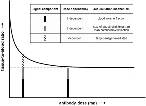
Ideally, monoclonal antibodies provide selective treatment by targeting the tumour, without affecting normal tissues. Therefore, antibody imaging is of interest, preferably in early stages of drug development. However, the imaging signal consists of specific, as well as non-specific, uptake. The aim of this study was to assess specific, target-mediated uptake in normal tissues, with immuno-PET in a phase I dose escalation study, using the anti-CD44 antibody RG7356 as example.
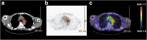
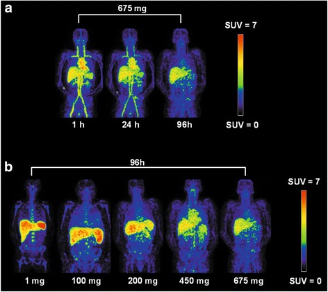
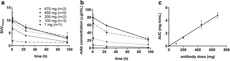
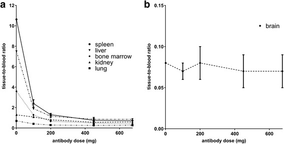
Data from thirteen patients with CD44-expressing solid tumours included in an imaging sub-study of a phase I dose escalation clinical trial using the anti-CD44 antibody RG7356 was analysed. Zirconium-labelled RG7356 (1 mg; 37 MBq) was administered after a variable dose of unlabelled RG7356 (0 to 675 mg). Tracer uptake in normal tissues (liver, spleen, kidney, lung, bone marrow, brain and blood pool) was used to calculate the area under the time antibody concentration curve (AUC) and expressed as tissue-to-blood AUC ratios. Within the dose range of 1 to 450 mg, tissue-to-blood AUC ratios decreased from 10.6 to 0.75 ± 0.16 for the spleen, 7.5 to 0.86 ± 0.18 for the liver, 3.6 to 0.48 ± 0.13 for the bone marrow, 0.69 to 0.26 ± 0.1 for the lung and 1.29 to 0.56 ± 0.14 for the kidney, indicating dose-dependent uptake. In all patients receiving ≥ 450 mg (n = 7), tumour uptake of the antibody was observed.
This study demonstrates how immuno-PET in a dose escalation study provides a non-invasive technique to quantify dose-dependent uptake in normal tissues, indicating specific, target-mediated uptake.
理想情况下,单克隆抗体通过靶向肿瘤提供选择性治疗,而不影响正常组织。因此,抗体成像备受关注,尤其在药物研发的早期阶段。然而,成像信号包括特异性摄取和非特异性摄取。本研究的目的是以抗CD44抗体RG7356为例,在I期剂量递增研究中利用免疫正电子发射断层扫描(immuno-PET)评估正常组织中特异性的、靶点介导的摄取。
分析了13例表达CD44的实体瘤患者的数据,这些患者纳入了一项使用抗CD44抗体RG7356的I期剂量递增临床试验的成像子研究。在给予不同剂量的未标记RG7356(0至675毫克)后,注射锆标记的RG7356(1毫克;37兆贝可)。利用正常组织(肝脏、脾脏、肾脏、肺、骨髓、脑和血池)中的示踪剂摄取来计算时间-抗体浓度曲线下面积(AUC),并表示为组织-血AUC比值。在1至450毫克的剂量范围内,脾脏的组织-血AUC比值从10.6降至0.75±0.16,肝脏从7.5降至0.86±0.18,骨髓从3.6降至0.48±0.13,肺从0.69降至0.26±0.1,肾脏从1.29降至0.56±0.14,表明存在剂量依赖性摄取。在所有接受≥450毫克(n = 7)的患者中,观察到了抗体在肿瘤中的摄取。
本研究证明了在剂量递增研究中免疫正电子发射断层扫描如何提供一种非侵入性技术来量化正常组织中的剂量依赖性摄取,表明存在特异性的、靶点介导的摄取。