Franchi Sira A, Macco Romina, Astro Veronica, Tonoli Diletta, Savino Elisa, Valtorta Flavia, Sala Kristyna, Botta Martina, de Curtis Ivan
Cell Adhesion Unit San Raffaele Scientific Institute and San Raffaele University, Milan, Italy.
Neuropsychopharmacology Unit, Division of Neuroscience, San Raffaele Scientific Institute and San Raffaele University, Milan, Italy.
Front Cell Neurosci. 2018 Jan 8;11:423. doi: 10.3389/fncel.2017.00423. eCollection 2017.
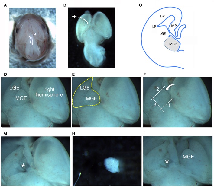
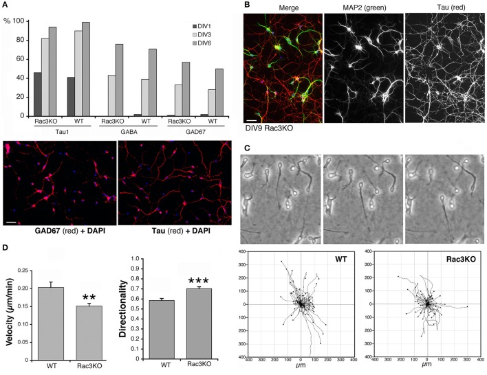
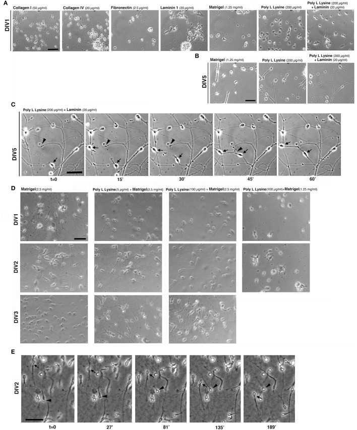
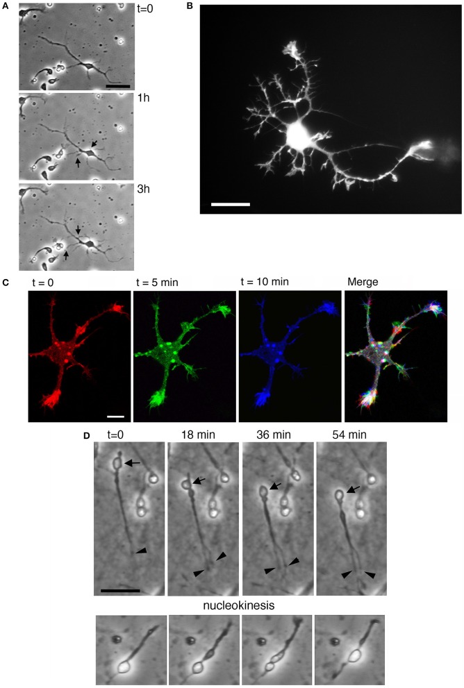
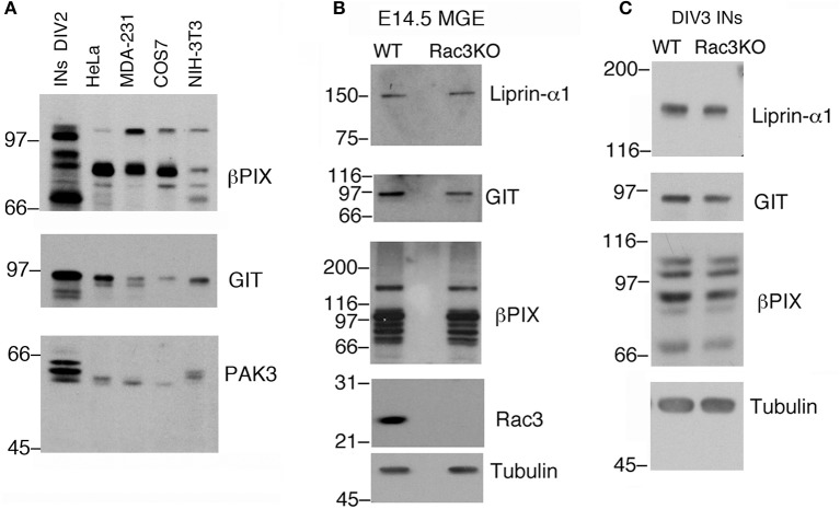
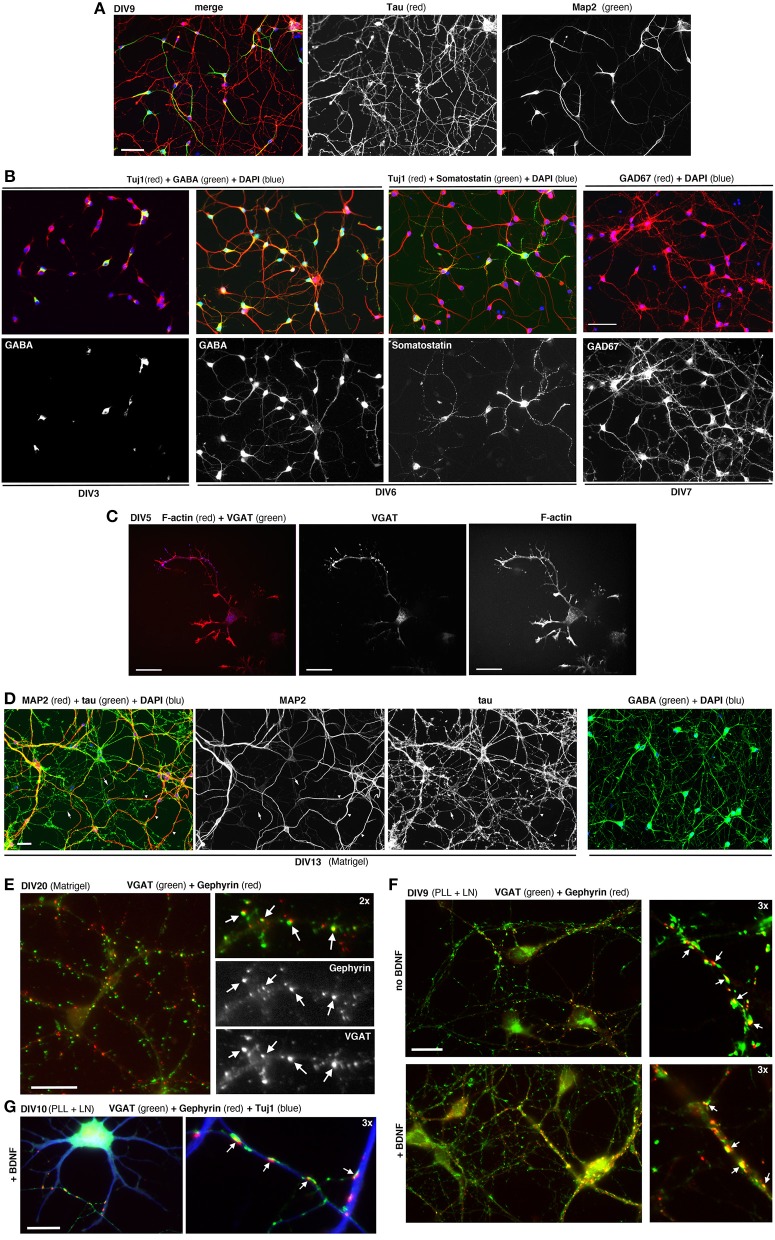
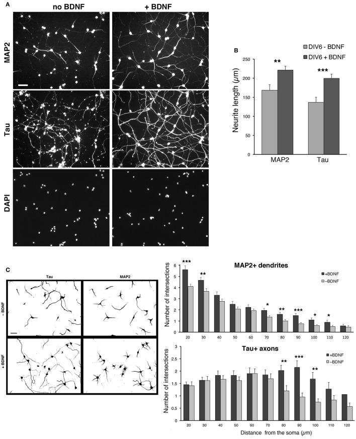
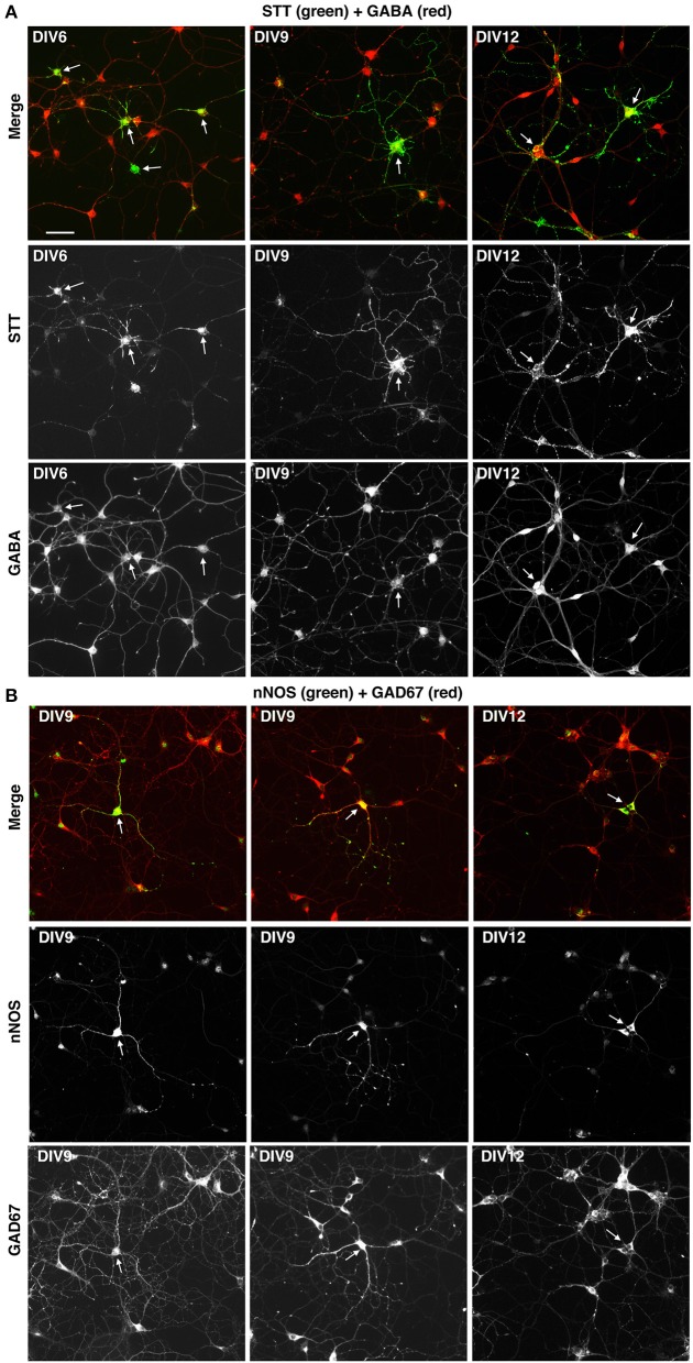
Understanding the mechanisms guiding interneuron development is a central aspect of the current research on cortical/hippocampal interneurons, which is highly relevant to brain function and pathology. In this methodological study we have addressed the setup of protocols for the reproducible culture of dissociated cells from murine medial ganglionic eminences (MGEs), to provide a culture system for the analysis of interneurons . This study includes the detailed protocols for the preparation of the dissociated cells, and for their culture on optimal substrates for cell migration or differentiation. These cultures enriched in interneurons may allow the investigation of the migratory behavior of interneuron precursors and their differentiation , up to the formation of morphologically identifiable GABAergic synapses. Live imaging of MGE-derived cells plated on proper substrates shows that they are useful to study the migratory behavior of the precursors, as well as the behavior of growth cones during the development of neurites. Most MGE-derived precursors develop into polarized GABAergic interneurons as determined by axonal, dendritic, and GABAergic markers. We present also a comparison of cells from WT and mutant mice as a proof of principle for the use of these cultures for the analysis of the migration and differentiation of GABAergic cells with different genetic backgrounds. The culture enriched in interneurons described here represents a useful experimental system to examine in a relatively easy and fast way the morpho-functional properties of these cells under physiological or pathological conditions, providing a powerful tool to complement the studies .
了解指导中间神经元发育的机制是当前皮质/海马中间神经元研究的核心内容,这与脑功能和病理学高度相关。在这项方法学研究中,我们探讨了从鼠内侧神经节隆起(MGEs)分离细胞进行可重复培养的方案设置,以提供一个用于分析中间神经元的培养系统。本研究包括分离细胞制备及其在细胞迁移或分化的最佳底物上培养的详细方案。这些富含中间神经元的培养物可能有助于研究中间神经元前体细胞的迁移行为及其分化,直至形成形态上可识别的γ-氨基丁酸能(GABAergic)突触。将MGE来源的细胞接种在合适底物上进行实时成像显示,它们有助于研究前体细胞的迁移行为以及神经突发育过程中生长锥的行为。根据轴突、树突和GABA能标记物判断,大多数MGE来源的前体细胞发育为极化的GABA能中间神经元。我们还比较了野生型和突变型小鼠的细胞,以此作为使用这些培养物分析不同遗传背景下GABA能细胞迁移和分化的原理证明。这里描述的富含中间神经元的培养物是一个有用的实验系统,可相对简便快速地检测这些细胞在生理或病理条件下的形态功能特性,并为补充相关研究提供有力工具。