Chen Chen, Zhang Yi Ping, Sun Yan, Xiong Wenhui, Shields Lisa B E, Shields Christopher B, Jin Xiaoming, Xu Xiao-Ming
Spinal Cord and Brain Injury Research Group, Stark Neurosciences Research Institute, and Department of Neurological Surgery, Indiana University School of Medicine; Program in Medical Neuroscience, Stark Neurosciences Research Institute, Indiana University School of Medicine.
Norton Neuroscience Institute, Norton Healthcare.
J Vis Exp. 2017 Dec 31(130):56565. doi: 10.3791/56565.
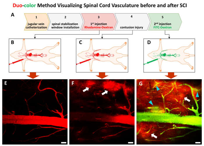
Spinal cord injury (SCI) causes significant vascular disruption at the site of injury. Vascular pathology occurs immediately after SCI and continues throughout the acute injury phase. In fact, endothelial cells appear to be the first to die after a contusive SCI. The early vascular events, including increased permeability of the blood-spinal cord barrier (BSCB), induce vasogenic edema and contribute to detrimental secondary injury events caused by complex injury mechanisms. Targeting the vascular disruption, therefore, could be a key strategy to reduce secondary injury cascades that contribute to histological and functional impairments after SCI. Previous studies were mostly performed on postmortem samples and were unable to capture the dynamic changes of the vascular network. In this study, we have developed an in vivo duo-color two-photon imaging method to monitor acute vascular dynamic changes following contusive SCI. This approach allows detecting blood flow, vessel diameter, and other vascular pathologies at various sites of the same rat pre- and post-injury. Overall, this method provides an excellent venue for investigating vascular dynamics.
脊髓损伤(SCI)会在损伤部位导致显著的血管破坏。血管病变在脊髓损伤后立即发生,并在整个急性损伤期持续存在。事实上,在挫伤性脊髓损伤后,内皮细胞似乎是最先死亡的细胞。早期血管事件,包括血脊髓屏障(BSCB)通透性增加,会诱发血管源性水肿,并导致由复杂损伤机制引起的有害继发性损伤事件。因此,针对血管破坏可能是减少继发性损伤级联反应的关键策略,这些级联反应会导致脊髓损伤后的组织学和功能障碍。以往的研究大多在尸检样本上进行,无法捕捉血管网络的动态变化。在本研究中,我们开发了一种体内双色双光子成像方法,以监测挫伤性脊髓损伤后的急性血管动态变化。这种方法可以在同一大鼠损伤前后的不同部位检测血流、血管直径和其他血管病变。总体而言,该方法为研究血管动力学提供了一个极佳的平台。