Department of Ophthalmology, Emory University, Atlanta, Georgia, United States.
Neuroscience, Emory University, Atlanta, Georgia, United States.
Invest Ophthalmol Vis Sci. 2018 Jan 1;59(1):572-581. doi: 10.1167/iovs.17-22692.
Electroretinograms (ERGs) are abnormal in diabetic retinas before the appearance of vascular lesions, providing a possible biomarker for diabetic vision loss. Previously, we reported that decreased retinal dopamine (DA) levels in diabetic rodents contributed to early visual and retinal dysfunction. In the current study, we examined whether oscillatory potentials (OPs) could serve as a potential marker for detecting early inner retinal dysfunction due to retinal DA deficiency.
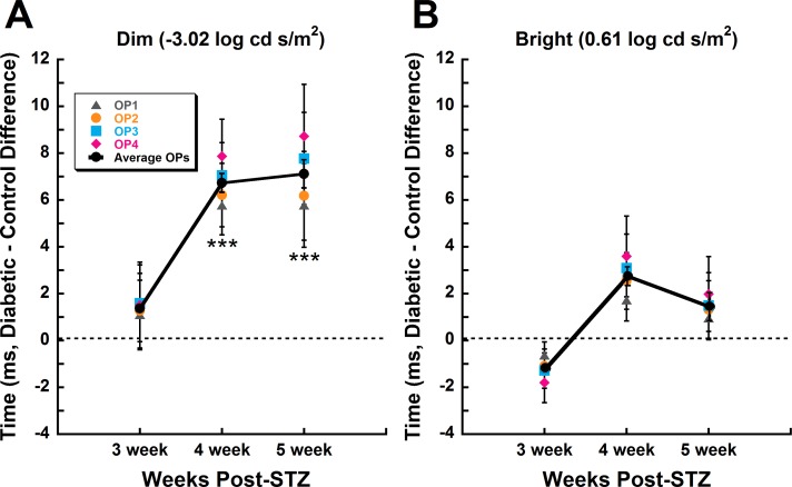
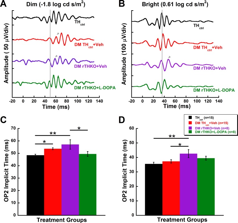
Retinal function was tested with dark-adapted ERGs, taken at 3, 4, and 5 weeks after diabetes induction with streptozotocin. Electrical responses were analyzed and correlations were made with previously reported retinal DA levels. The effect of restoring systemic DA levels or removing DA from the retina in diabetic mice on OPs was assessed using L-3,4-dihydroxyphenylalanine (L-DOPA) treatments and retina-specific tyrosine hydroxylase (Th) knockout mice (rTHKO), respectively.
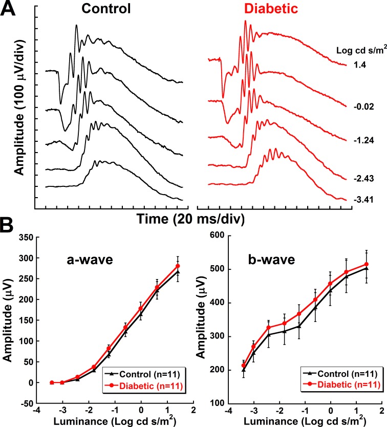
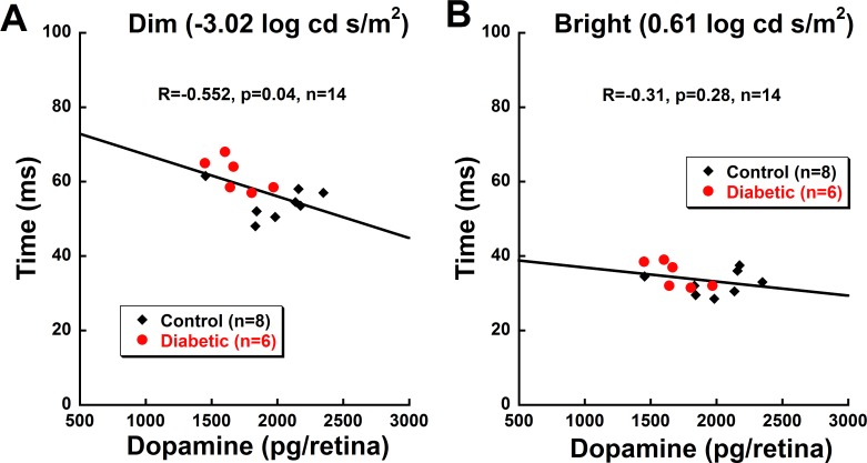
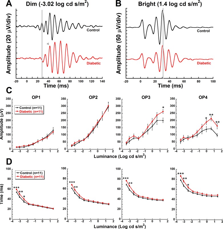
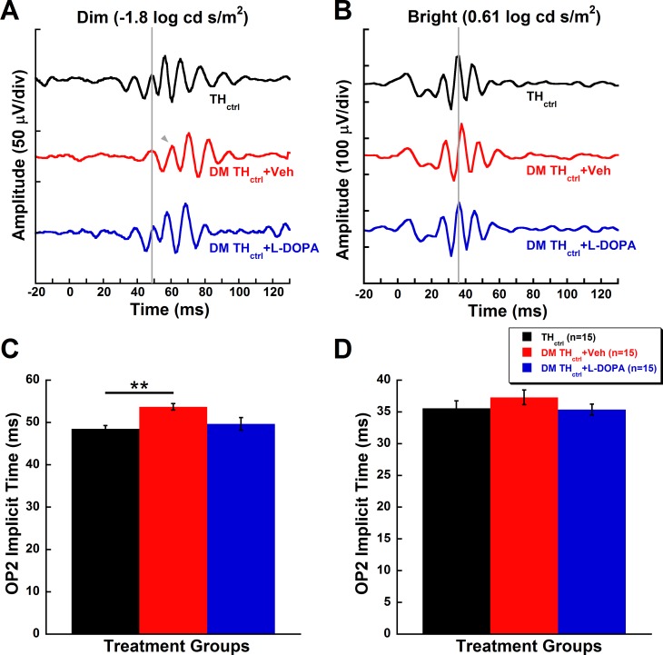
Diabetic animals had significantly delayed OPs compared to control animals in response to dim, but not bright, flash stimuli. L-DOPA treatment preserved OP implicit time in diabetic mice. Diabetic rTHKO mice had further delayed OPs compared to diabetic mice with normal retinal Th, with L-DOPA treatment also providing benefit. Decreasing retinal DA levels significantly correlated with increasing OP delays mediated by rod pathways.
Our data suggest that inner retinal dysfunction in early-stage diabetes is mediated by rod-pathway deficits and DA deficiencies. OP delays may be used to determine the earliest functional deficits in diabetic retinopathy and to establish an early treatment window for DA therapies that may prevent progressive vision loss.
在血管病变出现之前,糖尿病视网膜的视网膜电图(ERG)异常,为糖尿病视力丧失提供了一个可能的生物标志物。之前,我们报道了糖尿病啮齿动物的视网膜多巴胺(DA)水平降低导致早期视觉和视网膜功能障碍。在本研究中,我们研究了振荡电位(OP)是否可以作为检测由于视网膜 DA 缺乏引起的早期内视网膜功能障碍的潜在标志物。
用暗适应 ERG 在糖尿病诱导后 3、4 和 5 周检测视网膜功能。分析电响应,并与先前报道的视网膜 DA 水平进行相关性分析。使用 L-3,4-二羟基苯丙氨酸(L-DOPA)处理和视网膜特异性酪氨酸羟化酶(Th)敲除小鼠(rTHKO)分别评估恢复系统 DA 水平或从糖尿病小鼠中去除视网膜 DA 对 OP 的影响。
与对照动物相比,糖尿病动物对暗闪光刺激的 OP 潜伏期明显延迟,但对亮闪光刺激无明显延迟。L-DOPA 治疗可保留糖尿病小鼠的 OP 潜伏期。与具有正常视网膜 Th 的糖尿病小鼠相比,糖尿病 rTHKO 小鼠的 OP 潜伏期进一步延迟,L-DOPA 治疗也提供了益处。视网膜 DA 水平的降低与由杆状通路介导的 OP 延迟显著相关。
我们的数据表明,早期糖尿病中的内视网膜功能障碍是由杆状通路缺陷和 DA 缺乏介导的。OP 延迟可能用于确定糖尿病视网膜病变的最早功能缺陷,并为可能预防进行性视力丧失的 DA 治疗建立早期治疗窗口。