Lee Pureun-Haneul, Kim Byeong-Gon, Lee Sun-Hye, Leikauf George D, Jang An-Soo
1Division of Allergy and Respiratory Medicine, Department of Internal Medicine, Soonchunhyang University Bucheon Hospital, 170 Jomaru-ro, Wonmi-gu, Bucheon, Gyeonggi-do 420-767 South Korea.
2Department of Environmental and Occupational Health, Graduate School of Public Health, University of Pittsburgh, Pittsburgh, PA USA.
Proteome Sci. 2018 Jan 17;16:2. doi: 10.1186/s12953-017-0130-4. eCollection 2018.
Acrolein (allyl Aldehyde) as one of smoke irritant exacerbates chronic airway diseases and increased in sputum of patients with asthma and chronic obstructive lung disease. But underlying mechanism remains unresolved. The aim of study was to identify protein expression in human lung microvascular endothelial cells (HMVEC-L) exposed to acrolein.
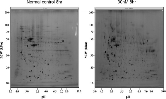
A proteomic approach was used to determine the different expression of proteins at 8 h and 24 h after treatment of acrolein 30 nM and 300 nM to HMVEC-L. Treatment of HMVEC-L with acrolein 30 nM and 300 nM altered 21 protein spots on the two-dimensional gel, and these were then analyzed by MALDI-TOF MS.
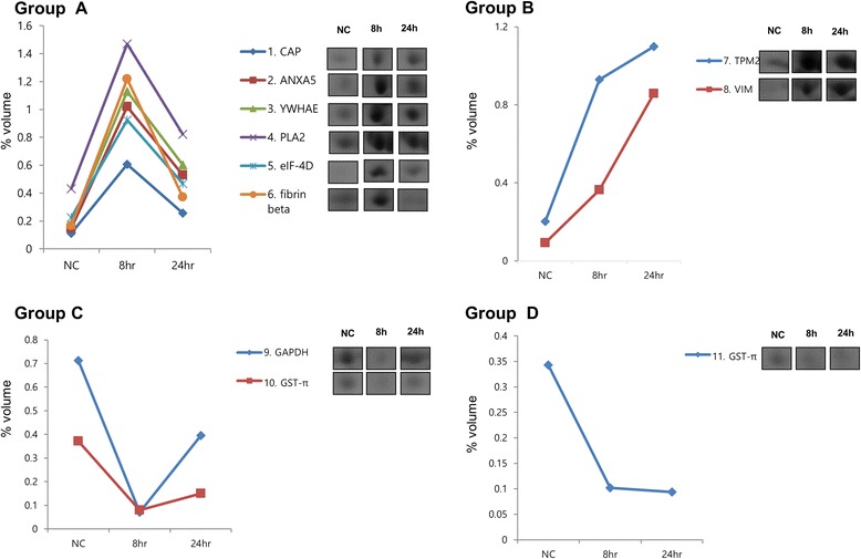
These proteins included antioxidant, signal transduction, cytoskeleton, protein transduction, catalytic reduction. The proteins were classified into four groups according to the time course of their expression patterns such as continually increasing, transient increasing, transient decreasing, and continually decreasing. For validation immunohistochemical staining and Western blotting was performed on lung tissues from acrolein exposed mice. Moesin was expressed in endothelium, epithelium, and inflammatory cells and increased in lung tissues of acrolein exposed mice compared with sham treated mice.
These results indicate that some of proteins may be an important role for airway disease exacerbation caused by acrolein exposure.
丙烯醛(烯丙醛)作为烟雾刺激物之一,会加重慢性气道疾病,且在哮喘和慢性阻塞性肺疾病患者的痰液中含量增加。但其潜在机制仍未明确。本研究的目的是确定暴露于丙烯醛的人肺微血管内皮细胞(HMVEC-L)中的蛋白质表达情况。
采用蛋白质组学方法,测定用30 nM和300 nM丙烯醛处理HMVEC-L 8小时和24小时后蛋白质的差异表达。用30 nM和300 nM丙烯醛处理HMVEC-L后,二维凝胶上有21个蛋白点发生改变,随后用基质辅助激光解吸电离飞行时间质谱(MALDI-TOF MS)进行分析。
这些蛋白质包括抗氧化剂、信号转导、细胞骨架、蛋白质转导、催化还原。根据其表达模式的时间进程,这些蛋白质被分为四组,即持续增加、短暂增加、短暂减少和持续减少。为进行验证,对暴露于丙烯醛的小鼠的肺组织进行了免疫组织化学染色和蛋白质印迹分析。肌动蛋白结合蛋白(Moesin)在内皮细胞、上皮细胞和炎性细胞中表达,与假处理小鼠相比,暴露于丙烯醛的小鼠肺组织中Moesin表达增加。
这些结果表明,某些蛋白质可能在丙烯醛暴露引起的气道疾病加重中起重要作用。