Abidoye Ibukun A, Ayoola Oluwagbemiga O, Idowu Bukunmi M, Aderibigbe Adeniyi S, Loto Olabisi M
Department of Radiology, Obafemi Awolowo University Teaching Hospitals Complex, Ile - Ife, Osun state, Nigeria.
Obstetrics and Gynecology, Obafemi Awolowo University Teaching Hospitals Complex, Ile - Ife, Osun state, Nigeria.
J Ultrason. 2017 Dec;17(71):253-258. doi: 10.15557/JoU.2017.0037. Epub 2017 Dec 29.
To evaluate the value of uterine artery Doppler indices and waveform pattern in predicting fetuses at risk for intrauterine growth restriction in hypertensive disorders of pregnancy.
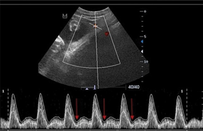
This was a prospective cross-sectional study including 80 pregnant subjects with hypertensive disorders of pregnancy and two control groups. Uterine artery Doppler sonography was performed in all study participants. Uterine artery Doppler indices across the groups were compared using the analysis of variance (ANOVA) while the presence of prediastolic notch was analyzed with the Chi Square test.
For the hypertensive disorders of pregnancy group, resistivity index > 0.66 had a sensitivity of 50.0%, specificity of 69.1% and a positive predictive value of 22.2% for predicting intrauterine growth restriction. The odds ratio was 2.2 with a 95% confidence interval of 0.6-7.8. The presence of prediastolic notching had a sensitivity of 100.0%, specificity of 96.0% and a positive predictive value of 80.0% for predicting intrauterine growth restriction. The odds ratio was 22.7 with a 95% confidence interval of 7.5-68.5.
Uterine artery Doppler sonography is useful for predicting fetuses at risk for intrauterine growth restriction in hypertensive disorder of pregnancy. Prediastolic notching is more sensitive and more specific than uterine artery resistivity index in predicting fetuses at risk of intrauterine growth restriction in established hypertensive disorder of pregnancy.
评估子宫动脉多普勒指数和波形模式在预测妊娠高血压疾病中存在宫内生长受限风险胎儿方面的价值。
这是一项前瞻性横断面研究,纳入80例患有妊娠高血压疾病的孕妇及两个对照组。对所有研究参与者进行子宫动脉多普勒超声检查。采用方差分析(ANOVA)比较各组间子宫动脉多普勒指数,采用卡方检验分析舒张前期切迹的存在情况。
对于妊娠高血压疾病组,阻力指数>0.66预测宫内生长受限的敏感度为50.0%,特异度为69.1%,阳性预测值为22.2%。比值比为2.2,95%置信区间为0.6 - 7.8。舒张前期切迹的存在预测宫内生长受限的敏感度为100.0%,特异度为96.0%,阳性预测值为80.0%。比值比为22.7,95%置信区间为7.5 - 68.5。
子宫动脉多普勒超声有助于预测妊娠高血压疾病中存在宫内生长受限风险的胎儿。在已确诊的妊娠高血压疾病中,舒张前期切迹在预测存在宫内生长受限风险胎儿方面比子宫动脉阻力指数更敏感、更特异。