Hofstoetter Ursula S, Freundl Brigitta, Binder Heinrich, Minassian Karen
Center for Medical Physics and Biomedical Engineering, Medical University Vienna, Vienna, Austria.
Neurological Center, Maria Theresien Schloessel, Otto Wagner Hospital, Vienna, Austria.
PLoS One. 2018 Jan 30;13(1):e0192013. doi: 10.1371/journal.pone.0192013. eCollection 2018.
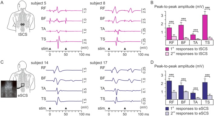
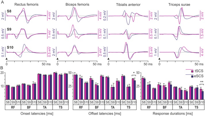
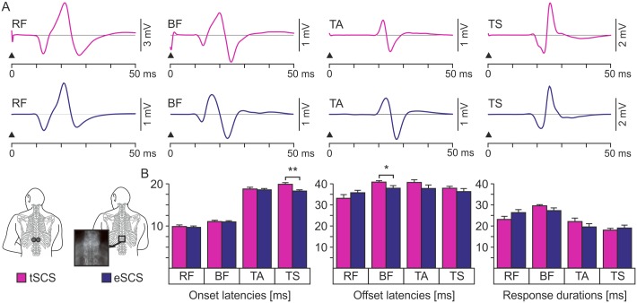
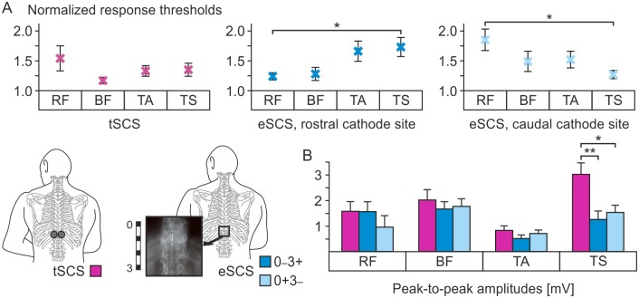
Epidural electrical stimulation of the lumbar spinal cord is currently regaining momentum as a neuromodulation intervention in spinal cord injury (SCI) to modify dysregulated sensorimotor functions and augment residual motor capacity. There is ample evidence that it engages spinal circuits through the electrical stimulation of large-to-medium diameter afferent fibers within lumbar and upper sacral posterior roots. Recent pilot studies suggested that the surface electrode-based method of transcutaneous spinal cord stimulation (SCS) may produce similar neuromodulatory effects as caused by epidural SCS. Neurophysiological and computer modeling studies proposed that this noninvasive technique stimulates posterior-root fibers as well, likely activating similar input structures to the spinal cord as epidural stimulation. Here, we add a yet missing piece of evidence substantiating this assumption. We conducted in-depth analyses and direct comparisons of the electromyographic (EMG) characteristics of short-latency responses in multiple leg muscles to both stimulation techniques derived from ten individuals with SCI each. Post-activation depression of responses evoked by paired pulses applied either epidurally or transcutaneously confirmed the reflex nature of the responses. The muscle responses to both techniques had the same latencies, EMG peak-to-peak amplitudes, and waveforms, except for smaller responses with shorter onset latencies in the triceps surae muscle group and shorter offsets of the responses in the biceps femoris muscle during epidural stimulation. Responses obtained in three subjects tested with both methods at different time points had near-identical waveforms per muscle group as well as same onset latencies. The present results strongly corroborate the activation of common neural input structures to the lumbar spinal cord-predominantly primary afferent fibers within multiple posterior roots-by both techniques and add to unraveling the basic mechanisms underlying electrical SCS.
作为一种神经调节干预手段,腰段脊髓硬膜外电刺激目前在脊髓损伤(SCI)治疗中重新获得关注,旨在改善失调的感觉运动功能并增强残余运动能力。有充分证据表明,它通过电刺激腰段和上骶段后根内的中到大直径传入纤维来激活脊髓回路。最近的初步研究表明,基于表面电极的经皮脊髓刺激(SCS)方法可能产生与硬膜外SCS相似的神经调节效果。神经生理学和计算机建模研究提出,这种非侵入性技术也会刺激后根纤维,可能激活与硬膜外刺激类似的脊髓输入结构。在此,我们补充了一项缺失的证据来支持这一假设。我们对来自10名脊髓损伤患者的两种刺激技术所引发的多条腿部肌肉短潜伏期反应的肌电图(EMG)特征进行了深入分析和直接比较。硬膜外或经皮施加配对脉冲所诱发反应的激活后抑制证实了这些反应的反射性质。两种技术引发的肌肉反应具有相同的潜伏期、EMG峰峰值幅度和波形,但在硬膜外刺激期间,比目鱼肌肌群的反应起始潜伏期较短且反应幅度较小,股二头肌的反应结束时间较短。在不同时间点接受两种方法测试的三名受试者中,每个肌肉群获得的反应具有几乎相同的波形以及相同的起始潜伏期。目前的结果有力地证实了两种技术均激活了腰段脊髓的共同神经输入结构——主要是多条后根内的初级传入纤维,并有助于揭示电刺激SCS的基本机制。