Izaz Shaik, Mandava Pragna, Bolla Nagesh, Dasari Bhargavi
Department of Conservative Dentistry and Endodontics, Sibar Institute of Dental Sciences, Guntur, Andhra Pradesh, India.
Department of Oral Medicine and Radiology, Sibar Institute of Dental Sciences, Guntur, Andhra Pradesh, India.
J Conserv Dent. 2017 Sep-Oct;20(5):370-373. doi: 10.4103/JCD.JCD_279_16.
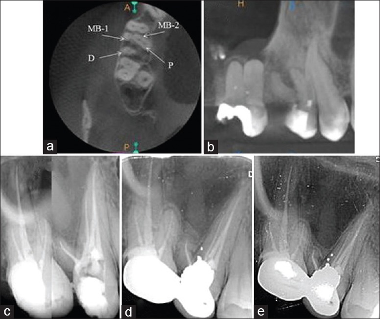
Knowledge and understanding the anatomical configuration of individual tooth play a significant role in success of endodontic treatment, in addition to through debridement and obturation of the canals. The canal anatomy of maxillary second premolar has been studied extensively, and the presence of a significant variety of multirooted canals is relatively rare in it. A 27-year-old female reported with a chief complaint of pain in her upper right posterior region for 10 days. On intraoral hard tissue examination, ill-defined access preparation was seen in maxillary right second premolar with exposed pulp. An intraoral periapical radiograph reveals radiolucency involving the pulp space and varied morphology in the same tooth. The occurrence of three roots with four canals in the maxillary second premolar is rare and not documented in the literature so far. This case report describes the nonsurgical endodontic management of such varied anatomical configuration using cone beam computed tomography as an evaluating diagnostic tool.
除了通过根管清创和充填外,了解和认识单颗牙齿的解剖结构在根管治疗的成功中起着重要作用。上颌第二前磨牙的根管解剖已得到广泛研究,其中存在多种多根管的情况相对少见。一名27岁女性主诉右上后牙区疼痛10天。口腔硬组织检查时,在上颌右第二前磨牙可见不明确的开髓预备且牙髓暴露。口腔根尖片显示牙髓腔有透射区且同一颗牙齿形态各异。上颌第二前磨牙出现三根四管的情况很罕见,目前文献中尚无记载。本病例报告描述了使用锥形束计算机断层扫描作为评估诊断工具,对这种复杂解剖结构进行非手术根管治疗的情况。