Schroeder Marion, Kjellström Ulrika
Lund University, Skane University Hospital, Department of Clinical Science Lund, Ophthalmology, Lund, Sweden.
Mol Vis. 2018 Jan 4;24:1-16. eCollection 2018.
To assess retinal function in combination with the retinal structure in -associated retinal degenerations. Moreover, to evaluate the possibility of predicting the natural course of these disorders.
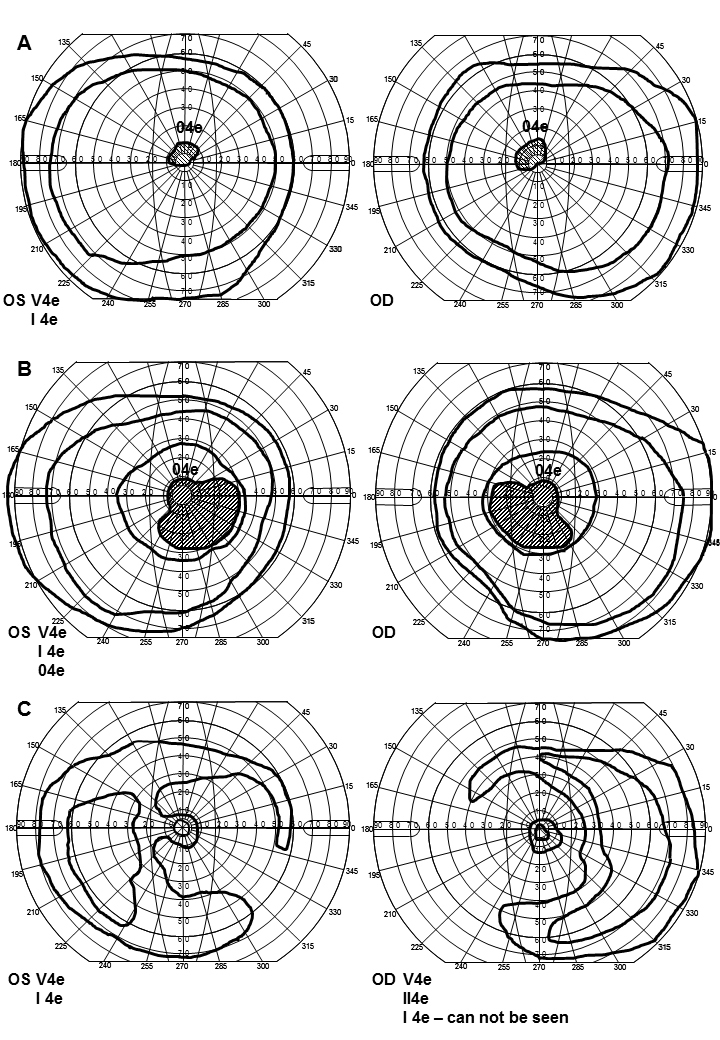
34 patients with Stargardt disease or cone rod dystrophy carrying confirmed mutations in were selected from our retinitis pigmentosa (RP) register. Sequence analysis of the entire coding region of the gene was performed. The patients were subdivided into three groups based on their most recent visual fields. Group 1 included ten patients with central scotomas within 10°, group 2 included 19 patients with larger central scotomas of 10-35°, and group 3 included five patients with mere temporal residues. The patients underwent slit-lamp and fundus examinations, visual acuity testing, optical coherence tomography (OCT), fundus photography (color, red-free, and autofluorescence (AF) images), full-field electroretinography (ffERG), and multifocal electroretinography (mERG). FfERG and mERG results were analyzed statistically. Total rod and cone function, as well as macular function, was compared between the three groups and of each group to a normal material. In 23 patients who had undergone ffERG on a previous occasion, the 30 Hz flicker implicit time (IT) from the first visit was also analyzed.
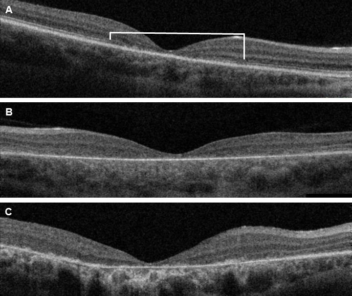
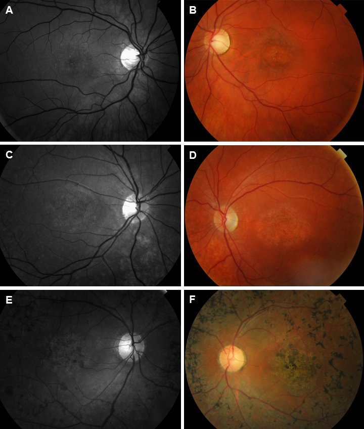
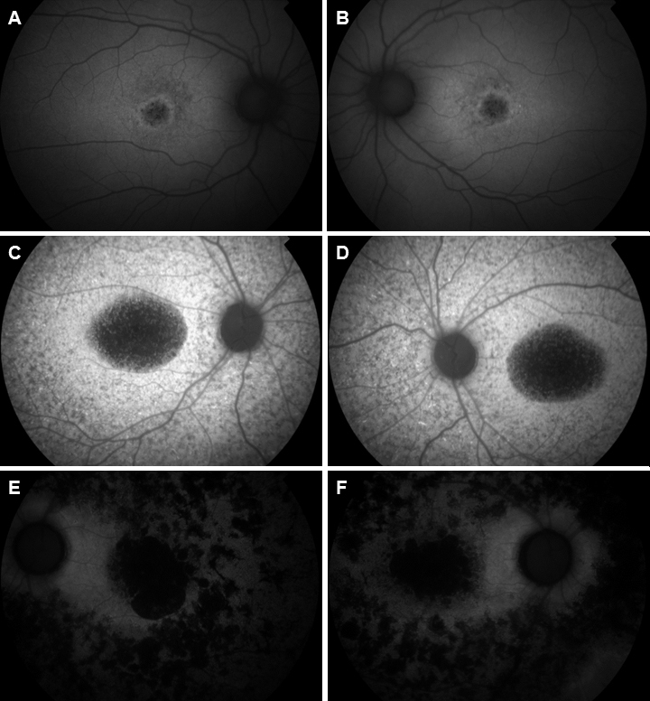
The ffERG statistics revealed significant differences between the groups regarding cone and rod function with group 1 showing the highest amplitudes and the shortest ITs while group 3 demonstrated the lowest amplitudes and the most delayed ITs. When compared to controls, group 1 did not show any significant changes while groups 2 and 3 demonstrated reduced amplitudes and delayed 30 Hz ITs. Regarding estimation of the natural course, identical results of the 30 Hz IT were encountered for the groups also at the first visit early in the course of disease. Comparison of the mERGs showed significant differences with group 1 demonstrating the highest amplitudes and group 3 the lowest for all rings but rings 2 and 3 in the right eye for which the amplitudes were the second highest. The mERGs for each group were also compared to controls showing reduced mERG amplitudes for all rings in all groups, except group 1, left eye. OCT showed macular attenuation in all patients. Evaluation of the inner and outer photoreceptor junction (IS/OS) morphology revealed alterations related to macular function measured with mERG in all eyes. Eight patients in group 1 showed foveal IS/OS junction loss, one had foveal IS/OS junction disorganization, and one had IS/OS loss also beyond the fovea. In group 2, one patient had IS/OS junction loss confined to the fovea, and the rest showed total loss of IS/OS junctions. Group 3 was devoid of IS/OS junctions. Concerning the AF images, group 1 showed small areas of absent AF in the macula, peripapillary sparing, and flecks of increased and reduced AF in the posterior pole. In group 2, the central areas of absent AF were larger. Flecks of reduced AF were the most dominant and reached beyond the posterior pole. Seven of 19 patients had peripapillary sparing. In group 3, large confluent areas of reduced AF were found in the posterior pole and beyond with small areas of increased AF in the far periphery. No peripapillary sparing was seen.
The current study demonstrates a significant difference in total retinal function, as well as macular function, between patients with -associated retinal degeneration and a different degree of visual field defects with gradual deterioration of function along with increased visual field constriction. Likewise, the morphological changes, including the deviant AF pattern and loss of IS/OS junctions, that were related to macular function measured with mERG worsened with the degree of visual field defects. Moreover, in these groups of patients with -associated retinal degenerations, full-field cone 30 Hz flicker IT seems to be a predictor of the natural course of the disease also on long-term follow-up.
评估与视网膜变性相关的视网膜功能及其结构。此外,评估预测这些疾病自然病程的可能性。
从我们的视网膜色素变性(RP)登记册中选取34例患有Stargardt病或锥杆营养不良且携带已确认突变的患者。对该基因的整个编码区进行序列分析。根据患者最近的视野将其分为三组。第1组包括10例中心暗点在10°以内的患者,第2组包括19例中心暗点较大(10 - 35°)的患者,第3组包括5例仅残留颞侧视野的患者。患者接受了裂隙灯和眼底检查、视力测试、光学相干断层扫描(OCT)、眼底照相(彩色、无赤光和自发荧光(AF)图像)、全视野视网膜电图(ffERG)和多焦视网膜电图(mERG)。对ffERG和mERG结果进行统计学分析。比较了三组之间以及每组与正常对照材料的总视杆和视锥功能以及黄斑功能。对23例之前接受过ffERG检查的患者,还分析了首次就诊时的30Hz闪烁潜伏时间(IT)。
ffERG统计显示,各组之间在视锥和视杆功能方面存在显著差异,第1组显示出最高的振幅和最短的IT,而第3组显示出最低的振幅和最延迟的IT。与对照组相比,第(1)组没有显示出任何显著变化,而第2组和第3组显示出振幅降低和30Hz IT延迟。关于自然病程的估计,在疾病早期的首次就诊时,各组的30Hz IT也得到了相同的结果。mERG的比较显示出显著差异,第1组在所有环上显示出最高的振幅,第3组除右眼的第2和第3环外,在所有环上显示出最低的振幅,而右眼的第2和第3环的振幅是第二高的。还将每组的mERG与对照组进行了比较,结果显示除第1组左眼外,所有组的所有环的mERG振幅均降低。OCT显示所有患者的黄斑均有萎缩。对内外光感受器连接(IS/OS)形态的评估显示,所有眼睛中与用mERG测量的黄斑功能相关的改变。第1组的8例患者显示中央凹IS/OS连接丧失,1例中央凹IS/OS连接紊乱,1例中央凹以外也有IS/OS丧失。在第2组中,1例患者的IS/OS连接丧失局限于中央凹,其余患者显示IS/OS连接完全丧失。第3组没有IS/OS连接。关于AF图像,第1组在黄斑区显示小面积的AF缺失,视乳头周围保留,后极有AF增加和减少的斑点。在第2组中,AF缺失的中央区域更大。AF减少的斑点最为突出,并延伸至后极以外。19例患者中有7例视乳头周围保留。在第3组中,后极及以外区域发现大片融合的AF减少区域,并在最外周有小面积的AF增加区域。未见视乳头周围保留。
当前研究表明,与视网膜变性相关的患者在总视网膜功能以及黄斑功能方面存在显著差异,随着视野缩小,功能逐渐恶化,视野缺损程度不同。同样,与用mERG测量的黄斑功能相关的形态学变化,包括异常的AF模式和IS/OS连接丧失,随着视野缺损程度的加重而恶化。此外,在这些与视网膜变性相关的患者组中,全视野视锥30Hz闪烁IT似乎也是疾病长期随访自然病程的一个预测指标。