Dobryakova Yulia V, Kasianov Artem, Zaichenko Maria I, Stepanichev Mikhail Y, Chesnokova Ekaterina A, Kolosov Petr M, Markevich Vladimir A, Bolshakov Alexey P
Institute of Higher Nervous Activity and Neurophysiology, Russian Academy of Sciences, Moscow, Russia.
Vavilov Institute of General Genetics, Russian Academy of Sciences, Moscow, Russia.
Front Mol Neurosci. 2018 Jan 17;10:429. doi: 10.3389/fnmol.2017.00429. eCollection 2017.
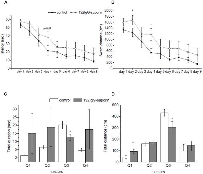
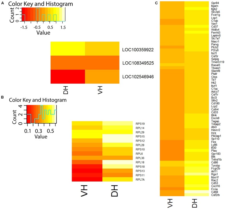
One of important aspects of development of Alzheimer's disease is degeneration of septal cholinergic neurons that innervate the hippocampus. We took advantage of widely used model of cholinergic deficit in the hippocampus, intracerebroventricular administration of IgG-saporin (Ig-saporin), to analyze the postponed consequences of cholinergic deficit in different parts of the hippocampus. We studied effects of the immunotoxin on the behavior of rats and gene expression in the dorsal and ventral hippocampus using RNA-seq approach. We found that under normal conditions dorsal and ventral parts of the hippocampus differ in the expression of 1129 protein-coding genes and 49 non-coding RNAs (ncRNAs) and do not differ in the expression of 10 microRNAs, which were detected in both parts of the hippocampus. Ig-saporin-induced degeneration of cholinergic septal neurons did not affect rat behavior in open field, T-maze, and passive avoidance task but impaired memory retention in Morris water maze. To analyze Ig-saporin-induced changes in the gene expression, we formed the following groups of genes: genes expressed exclusively in certain cell types (neurons, astrocytes, microglia, oligodendrocytes, and vascular cells) and, among universally expressed genes, a group of genes that encode ribosome-forming proteins. For all groups of genes, the alterations in the gene expression produced by the immunotoxin were stronger in the dorsal as compared to the ventral hippocampus. We found that, among groups of universally expressed genes, Ig-saporin increased the expression of ribosome-forming proteins in both dorsal and ventral hippocampus. Ig-saporin also strongly upregulated expression of microglia-specific genes only in the dorsal hippocampus. A subset of affected microglial genes comprised genes associated with inflammation, however, did not include genes related to acute inflammation such as interleukins-1b, -6, -15, and -18 as well as TNF. The expression of other cell-specific genes (genes specific for neurons, astrocytes, oligodendrocytes, and vascular cells) was unaffected. The data obtained suggest that disturbance of memory-associated behavior after administration of Ig-saporin is associated with upregulation of microglia-associated genes in the dorsal but not ventral hippocampus.
阿尔茨海默病发展的一个重要方面是支配海马体的隔区胆碱能神经元的退化。我们利用广泛使用的海马体胆碱能缺陷模型,即脑室内注射IgG-皂草素(Ig-皂草素),来分析海马体不同部位胆碱能缺陷的延迟后果。我们使用RNA测序方法研究了免疫毒素对大鼠行为以及背侧和腹侧海马体基因表达的影响。我们发现,在正常条件下,海马体的背侧和腹侧部分在1129个蛋白质编码基因和49个非编码RNA(ncRNA)的表达上存在差异,而在海马体两个部分均检测到的10个微小RNA的表达上没有差异。Ig-皂草素诱导的隔区胆碱能神经元退化在旷场、T迷宫和被动回避任务中不影响大鼠行为,但损害了莫里斯水迷宫中的记忆保持。为了分析Ig-皂草素诱导的基因表达变化,我们形成了以下几组基因:仅在某些细胞类型(神经元、星形胶质细胞、小胶质细胞、少突胶质细胞和血管细胞)中表达的基因,以及在普遍表达的基因中,一组编码核糖体形成蛋白的基因。对于所有基因组,与腹侧海马体相比,免疫毒素在背侧海马体中产生的基因表达变化更强。我们发现,在普遍表达的基因组中,Ig-皂草素增加了背侧和腹侧海马体中核糖体形成蛋白的表达。Ig-皂草素还仅在背侧海马体中强烈上调小胶质细胞特异性基因的表达。受影响的小胶质细胞基因的一个子集包括与炎症相关的基因,然而,不包括与急性炎症相关的基因,如白细胞介素-1β、-6、-15和-18以及肿瘤坏死因子。其他细胞特异性基因(神经元、星形胶质细胞、少突胶质细胞和血管细胞特异性基因)的表达未受影响。获得的数据表明,注射Ig-皂草素后记忆相关行为的紊乱与背侧而非腹侧海马体中小胶质细胞相关基因的上调有关。