• 文献检索
  • 文档翻译
  • 深度研究
  • 学术资讯
  • Suppr Zotero 插件Zotero 插件
  • 邀请有礼
  • 套餐&价格
  • 历史记录
应用&插件
Suppr Zotero 插件Zotero 插件浏览器插件Mac 客户端Windows 客户端微信小程序
定价
高级版会员购买积分包购买API积分包
服务
文献检索文档翻译深度研究API 文档MCP 服务
关于我们
关于 Suppr公司介绍联系我们用户协议隐私条款
关注我们

Suppr 超能文献

核心技术专利:CN118964589B侵权必究
粤ICP备2023148730 号-1Suppr @ 2026

文献检索

告别复杂PubMed语法,用中文像聊天一样搜索,搜遍4000万医学文献。AI智能推荐,让科研检索更轻松。

立即免费搜索

文件翻译

保留排版,准确专业,支持PDF/Word/PPT等文件格式,支持 12+语言互译。

免费翻译文档

深度研究

AI帮你快速写综述,25分钟生成高质量综述,智能提取关键信息,辅助科研写作。

立即免费体验

假性进展、放射性坏死及脑肿瘤复发的诊断与治疗

The diagnosis and treatment of pseudoprogression, radiation necrosis and brain tumor recurrence.

作者信息

Parvez Kashif, Parvez Aatif, Zadeh Gelareh

机构信息

Division of Neurosurgery, University Health Network, Toronto Western Hospital, University of Toronto, Toronto, ON M5T 2S8, Canada.

Department of Medical Imaging, Royal University Hospital, University of Saskatchewan, Saskatoon, SK S7N 0W8, Canada.

出版信息

Int J Mol Sci. 2014 Jul 3;15(7):11832-46. doi: 10.3390/ijms150711832.

DOI:10.3390/ijms150711832
PMID:24995696
原文链接:https://pmc.ncbi.nlm.nih.gov/articles/PMC4139817/
Abstract

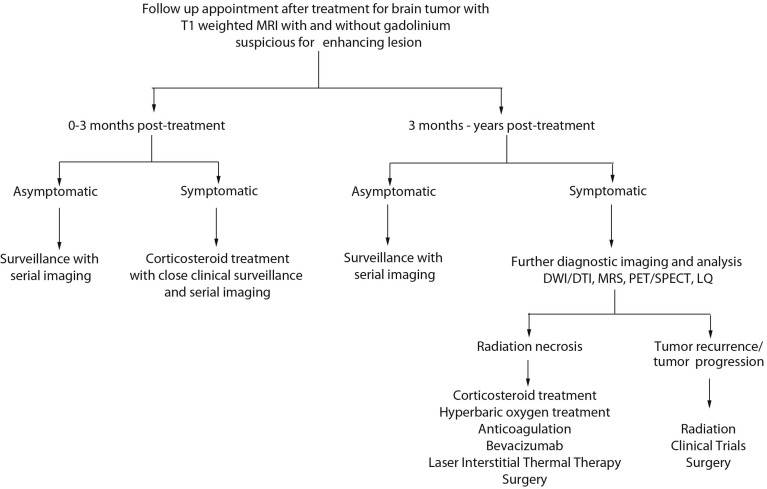
Radiation therapy is an important modality used in the treatment of patients with brain metastatic disease and malignant gliomas. Post-treatment surveillance often involves serial magnetic resonance imaging. A challenge faced by clinicians is in the diagnosis and management of a suspicious gadolinium-enhancing lesion found on imaging. The suspicious lesion may represent post-treatment radiation effects (PTRE) such as pseudoprogression, radiation necrosis or tumor recurrence. Significant progress has been made in diagnostic imaging modalities to assist in differentiating these entities. Surgical and medical interventions have also been developed to treat PTRE. In this review, we discuss the pathophysiology, clinical presentation, diagnostic imaging modalities and provide an algorithm for the management of pseudoprogression, radiation necrosis and tumor recurrence.

摘要

放射治疗是用于治疗脑转移瘤和恶性胶质瘤患者的一种重要治疗方式。治疗后的监测通常包括系列磁共振成像检查。临床医生面临的一个挑战是对影像学检查中发现的可疑钆增强病变进行诊断和管理。该可疑病变可能代表治疗后放射效应(PTRE),如假性进展、放射性坏死或肿瘤复发。在辅助鉴别这些病变的诊断成像方式方面已取得显著进展。也已开发出手术和药物干预措施来治疗PTRE。在本综述中,我们讨论其病理生理学、临床表现、诊断成像方式,并提供一个用于管理假性进展、放射性坏死和肿瘤复发的算法。

https://cdn.ncbi.nlm.nih.gov/pmc/blobs/fa2b/4139817/f9a39748b1bf/ijms-15-11832-g001.jpg

相似文献

1
The diagnosis and treatment of pseudoprogression, radiation necrosis and brain tumor recurrence.假性进展、放射性坏死及脑肿瘤复发的诊断与治疗
Int J Mol Sci. 2014 Jul 3;15(7):11832-46. doi: 10.3390/ijms150711832.
2
Radiation necrosis or glioma recurrence: is computer-assisted stereotactic biopsy useful?放射性坏死还是胶质瘤复发:计算机辅助立体定向活检有用吗?
J Neurosurg. 1995 Mar;82(3):436-44. doi: 10.3171/jns.1995.82.3.0436.
3
Distinguishing glioma recurrence from treatment effect after radiochemotherapy and immunotherapy.区分放射化疗和免疫治疗后胶质瘤复发与治疗效果。
Neurosurg Clin N Am. 2010 Jan;21(1):181-6. doi: 10.1016/j.nec.2009.08.003.
4
Treatment-related change versus tumor recurrence in high-grade gliomas: a diagnostic conundrum--use of dynamic susceptibility contrast-enhanced (DSC) perfusion MRI.高级别胶质瘤中的治疗相关性变化与肿瘤复发:诊断难题——动态磁敏感对比增强(DSC)灌注 MRI 的应用。
AJR Am J Roentgenol. 2012 Jan;198(1):19-26. doi: 10.2214/AJR.11.7417.
5
Discriminating radiation necrosis from tumor progression in gliomas: a systematic review what is the best imaging modality?鉴别脑胶质瘤中的放射性坏死与肿瘤进展:哪种影像学方法最佳?系统综述
J Neurooncol. 2013 Apr;112(2):141-52. doi: 10.1007/s11060-013-1059-9. Epub 2013 Jan 24.
6
Glioma recurrence versus radiation necrosis? A pilot comparison of arterial spin-labeled, dynamic susceptibility contrast enhanced MRI, and FDG-PET imaging.胶质瘤复发与放射性坏死?动脉自旋标记、动态磁敏感对比增强 MRI 和 FDG-PET 成像的初步比较。
Acad Radiol. 2010 Mar;17(3):282-90. doi: 10.1016/j.acra.2009.10.024. Epub 2010 Jan 12.
7
Radiation necrosis versus glioma recurrence: conventional MR imaging clues to diagnosis.放射性坏死与胶质瘤复发:传统磁共振成像的诊断线索
AJNR Am J Neuroradiol. 2005 Sep;26(8):1967-72.
8
Diffusion-weighted imaging in the follow-up of treated high-grade gliomas: tumor recurrence versus radiation injury.弥散加权成像在高级别胶质瘤治疗后的随访中:肿瘤复发与放射性损伤
AJNR Am J Neuroradiol. 2004 Feb;25(2):201-9.
9
Role of circulating tumor cell detection in differentiating tumor recurrence from treatment necrosis of brain gliomas.循环肿瘤细胞检测在区分脑胶质瘤肿瘤复发与治疗性坏死中的作用。
Biosci Trends. 2021 May 11;15(2):107-117. doi: 10.5582/bst.2021.01017. Epub 2021 Apr 29.
10
Progress in neuro-oncology: imaging in-phase!神经肿瘤学进展:影像学同步发展!
Diagn Interv Imaging. 2014 Oct;95(10):905. doi: 10.1016/j.diii.2014.09.001.

引用本文的文献

1
Auxiliary Value of [F]F-Fluorocholine PET/CT in Evaluating Post-Stereotactic Radiosurgery Recurrence of Lung Cancer Brain Metastases: A Comparative Analysis with Contrast-Enhanced MRI.[F]F-氟胆碱PET/CT在评估肺癌脑转移立体定向放射治疗后复发中的辅助价值:与对比增强MRI的对比分析
Cancers (Basel). 2025 Aug 7;17(15):2591. doi: 10.3390/cancers17152591.
2
Spatial Omics Profiling of Treatment-Naïve Lung Adenocarcinoma with Brain Metastasis as the Initial Presentation.以脑转移为首发表现的未经治疗的肺腺癌的空间组学分析
Cancers (Basel). 2025 Jul 31;17(15):2529. doi: 10.3390/cancers17152529.
3
Development and validation of a deep learning algorithm for discriminating glioma recurrence from radiation necrosis on MRI.

本文引用的文献

1
Inflammation as well as angiogenesis may participate in the pathophysiology of brain radiation necrosis.炎症以及血管生成可能参与脑放射性坏死的病理生理学过程。
J Radiat Res. 2014 Jul;55(4):803-11. doi: 10.1093/jrr/rru017. Epub 2014 Mar 27.
2
Extent of perilesional edema differentiates radionecrosis from tumor recurrence following stereotactic radiosurgery for brain metastases.瘤周水肿程度可区分脑转移瘤立体定向放射外科治疗后放射性坏死与肿瘤复发。
Neuro Oncol. 2013 Dec;15(12):1732-8. doi: 10.1093/neuonc/not130.
3
(18)F-FDOPA PET for differentiating recurrent or progressive brain metastatic tumors from late or delayed radiation injury after radiation treatment.
一种用于在磁共振成像(MRI)上鉴别胶质瘤复发与放射性坏死的深度学习算法的开发与验证
Front Oncol. 2025 Jun 6;15:1573700. doi: 10.3389/fonc.2025.1573700. eCollection 2025.
4
A twin-tower model using MRI and gene for prediction on brain tumor patients' response to therapy.一种使用磁共振成像(MRI)和基因的双塔模型,用于预测脑肿瘤患者对治疗的反应。
Bioinform Adv. 2025 Mar 4;5(1):vbaf041. doi: 10.1093/bioadv/vbaf041. eCollection 2025.
5
The diagnostic value of advanced tracer kinetic models in evaluating high grade gliomas recurrence and treatment response using dynamic contrast-enhanced MRI.先进示踪剂动力学模型在利用动态对比增强磁共振成像评估高级别胶质瘤复发及治疗反应中的诊断价值
Front Oncol. 2025 Apr 16;15:1536122. doi: 10.3389/fonc.2025.1536122. eCollection 2025.
6
Brain MRI and regional vulnerabilities to radiation necrosis: investigating the impact of stereotactic radiotherapy in brain metastases treatment.脑磁共振成像与放射性坏死的区域易损性:探究立体定向放射治疗在脑转移瘤治疗中的影响
Front Radiol. 2025 Apr 9;5:1554017. doi: 10.3389/fradi.2025.1554017. eCollection 2025.
7
Prediction of brain metastasis progression after stereotactic radiosurgery: sensitivity to changing the definition of progression.立体定向放射外科治疗后脑转移进展的预测:对进展定义变化的敏感性
J Med Imaging (Bellingham). 2025 Mar;12(2):024504. doi: 10.1117/1.JMI.12.2.024504. Epub 2025 Apr 8.
8
Intravoxel incoherent motion imaging and dynamic susceptibility contrast perfusion MRI in differentiation between recurrent intracranial tumor and treatment-induced changes.体素内不相干运动成像及动态磁敏感对比灌注磁共振成像在复发性颅内肿瘤与治疗后改变鉴别中的应用
Neuroradiology. 2025 Mar 21. doi: 10.1007/s00234-025-03575-4.
9
Comparison of Imaging Modalities in Differentiating Cerebral Neoplastic Lesions and Post-radiation Necrosis.鉴别脑肿瘤性病变与放射性坏死的成像模态比较
Cureus. 2025 Feb 6;17(2):e78653. doi: 10.7759/cureus.78653. eCollection 2025 Feb.
10
A Radiologist's Guide to IDH-Wildtype Glioblastoma for Efficient Communication With Clinicians: Part II-Essential Information on Post-Treatment Imaging.放射科医生与临床医生有效沟通的异柠檬酸脱氢酶野生型胶质母细胞瘤指南:第二部分——治疗后影像学的重要信息
Korean J Radiol. 2025 Apr;26(4):368-389. doi: 10.3348/kjr.2024.0983. Epub 2025 Jan 10.
(18)F-FDOPA PET 用于区分放疗后晚期或延迟性放射性损伤与复发性或进展性脑转移瘤。
J Nucl Med. 2014 Jan;55(1):30-6. doi: 10.2967/jnumed.113.121418. Epub 2013 Oct 28.
4
Differentiating tumor recurrence from treatment necrosis: a review of neuro-oncologic imaging strategies.肿瘤复发与治疗坏死的鉴别:神经肿瘤影像学策略综述。
Neuro Oncol. 2013 May;15(5):515-34. doi: 10.1093/neuonc/nos307. Epub 2013 Jan 16.
5
Evaluation of dynamic contrast-enhanced T1-weighted perfusion MRI in the differentiation of tumor recurrence from radiation necrosis.评估动态对比增强 T1 加权灌注 MRI 在肿瘤复发与放射性坏死鉴别中的应用。
Neuroradiology. 2013 Feb;55(3):361-9. doi: 10.1007/s00234-012-1127-4. Epub 2012 Dec 22.
6
Radiation necrosis in the brain: imaging features and differentiation from tumor recurrence.脑内放射性坏死:影像学特征与肿瘤复发的鉴别。
Radiographics. 2012 Sep-Oct;32(5):1343-59. doi: 10.1148/rg.325125002.
7
Laser interstitial thermal therapy for focal cerebral radiation necrosis: a case report and literature review.激光间质热疗治疗局灶性脑放射性坏死:一例报告及文献综述
Stereotact Funct Neurosurg. 2012;90(3):192-200. doi: 10.1159/000338251. Epub 2012 Jun 5.
8
Conventional MRI does not reliably distinguish radiation necrosis from tumor recurrence after stereotactic radiosurgery.常规 MRI 无法可靠地区分立体定向放射手术后的放射性坏死与肿瘤复发。
J Neurooncol. 2012 Aug;109(1):149-58. doi: 10.1007/s11060-012-0881-9. Epub 2012 May 26.
9
Radiation necrosis: relevance with respect to treatment of primary and secondary brain tumors.放射性坏死:与原发性和继发性脑肿瘤治疗的相关性。
Curr Neurol Neurosci Rep. 2012 Jun;12(3):276-85. doi: 10.1007/s11910-012-0258-7.
10
Radiation necrosis following treatment of high grade glioma--a review of the literature and current understanding.高级别脑胶质瘤治疗后放射性坏死——文献复习与目前认识。
Acta Neurochir (Wien). 2012 Feb;154(2):191-201; discussion 201. doi: 10.1007/s00701-011-1228-6. Epub 2011 Dec 1.