Department of Nursing and Department of Optometry, Hsin Sheng Junior College of Medical Care and Management, Taoyuan City, Taiwan.
Graduate Institute of Medical Sciences, College of Medicine, Taipei Medical University, Taipei, Taiwan.
Sci Rep. 2018 Feb 5;8(1):2368. doi: 10.1038/s41598-018-19654-x.
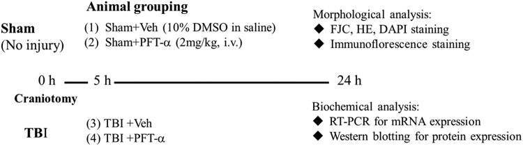
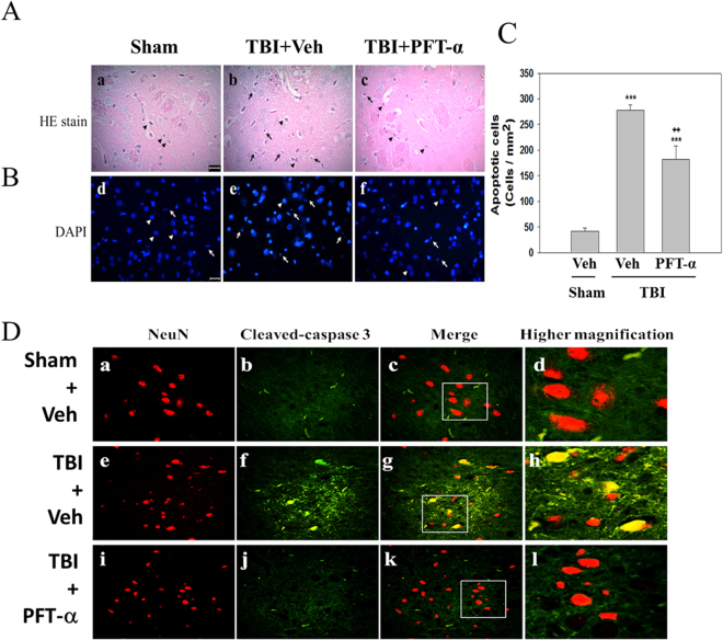
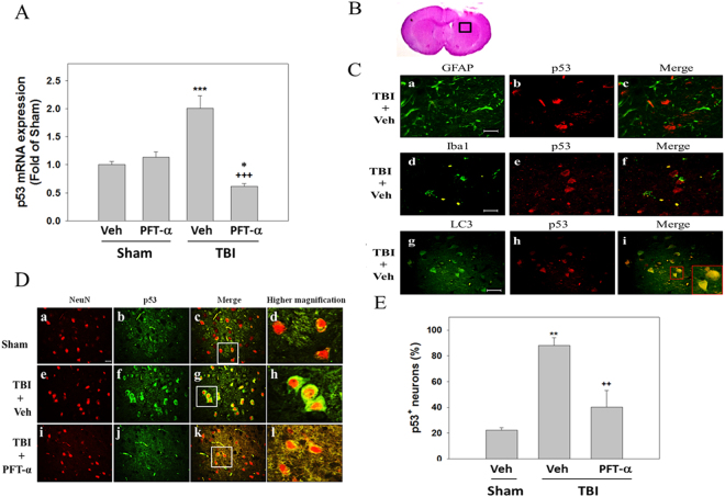
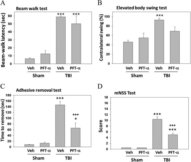
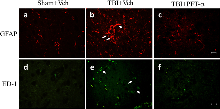
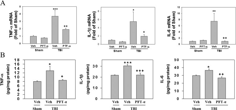
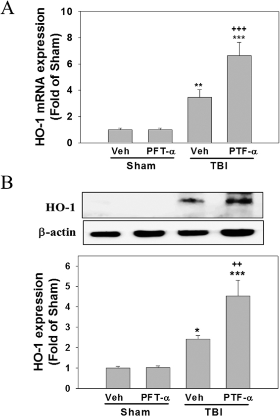
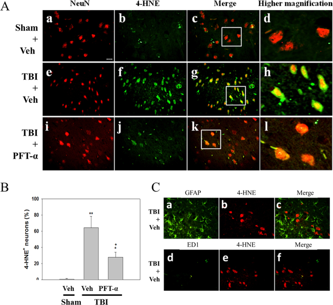
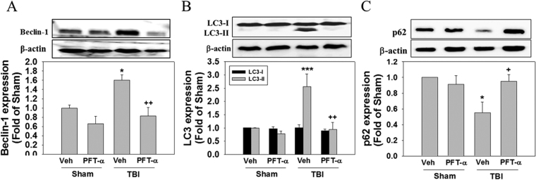
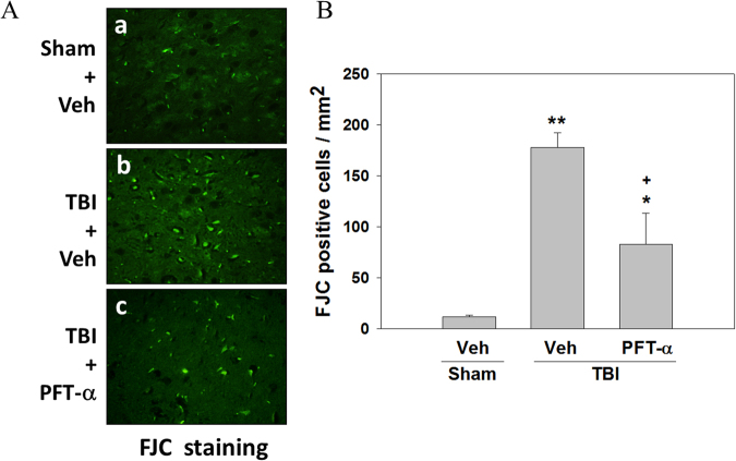
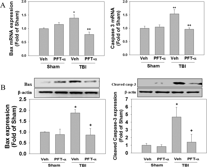
Cortical and hippocampal neuronal damages caused by traumatic brain injury (TBI) are associated with motor and cognitive impairments; however, only little attention paid to the striatal damage. It is known that the p53 tumor-suppressor transcription factor participated in TBI-induced secondary brain damage. We investigated how the p53 inactivator pifithrin (PFT)-α affected TBI-induced striatal neuronal damage at 24 h post-injury. Sprague-Dawley rats subjected to a controlled cortical impact were used as TBI models. We observed that p53 mRNA significantly increased, whereas p53 protein expression was distributed predominantly in neurons but not in glia cells in striatum after TBI. PFT-α improved motor deficit following TBI. PFT-α suppressed TBI-induced striatal glial activation and expression of proinflammatory cytokines. PFT-α alleviated TBI-induced oxidative damage TBI induced autophagy was evidenced by increased protein expression of Beclin-1 and shift of microtubule-associated light chain (LC)3-I to LC3-II, and decreased p62. These effects were reduced by PFT-α. Post-injury PFT-α treatment reduced the number of degenerating (FJC-positive) and apoptotic neurons. Our results suggest that PFT-α may provide neuroprotective effects via p53-dependent or -independent mechanisms depending on the cell type and timing after the TBI and can possibly be developed into a novel therapy to ameliorate TBI-induced neuronal damage.
创伤性脑损伤 (TBI) 引起的皮质和海马神经元损伤与运动和认知障碍有关;然而,只有很少的注意力集中在纹状体损伤上。已知 p53 肿瘤抑制转录因子参与 TBI 引起的继发性脑损伤。我们研究了 p53 失活剂 pifithrin-α (PFT-α) 在 TBI 后 24 小时如何影响 TBI 引起的纹状体神经元损伤。使用控制性皮质撞击的 Sprague-Dawley 大鼠作为 TBI 模型。我们观察到,TBI 后 p53 mRNA 显著增加,而 p53 蛋白表达主要分布在纹状体神经元中,而不是在胶质细胞中。PFT-α 改善了 TBI 后的运动障碍。PFT-α 抑制了 TBI 诱导的纹状体神经胶质激活和促炎细胞因子的表达。PFT-α 减轻了 TBI 诱导的氧化损伤 TBI 诱导的自噬通过增加 Beclin-1 蛋白表达和微管相关轻链 (LC)3-I 向 LC3-II 的转移以及 p62 的减少来证明。这些作用被 PFT-α 降低。损伤后 PFT-α 治疗减少了变性 (FJC 阳性) 和凋亡神经元的数量。我们的结果表明,PFT-α 可能通过 p53 依赖性或非依赖性机制提供神经保护作用,具体取决于 TBI 后细胞类型和时间,并可能开发成一种新的治疗方法来改善 TBI 引起的神经元损伤。