• 文献检索
  • 文档翻译
  • 深度研究
  • 学术资讯
  • Suppr Zotero 插件Zotero 插件
  • 邀请有礼
  • 套餐&价格
  • 历史记录
应用&插件
Suppr Zotero 插件Zotero 插件浏览器插件Mac 客户端Windows 客户端微信小程序
定价
高级版会员购买积分包购买API积分包
服务
文献检索文档翻译深度研究API 文档MCP 服务
关于我们
关于 Suppr公司介绍联系我们用户协议隐私条款
关注我们

Suppr 超能文献

核心技术专利:CN118964589B侵权必究
粤ICP备2023148730 号-1Suppr @ 2026

文献检索

告别复杂PubMed语法,用中文像聊天一样搜索,搜遍4000万医学文献。AI智能推荐,让科研检索更轻松。

立即免费搜索

文件翻译

保留排版,准确专业,支持PDF/Word/PPT等文件格式,支持 12+语言互译。

免费翻译文档

深度研究

AI帮你快速写综述,25分钟生成高质量综述,智能提取关键信息,辅助科研写作。

立即免费体验

在小鼠纤维化肝脏中由Jagged1/Notch2信号诱导的新型甲胎蛋白表达细胞群的鉴定

Identification of a novel alpha-fetoprotein-expressing cell population induced by the Jagged1/Notch2 signal in murine fibrotic liver.

作者信息

Nakano Yasuhiro, Nakao Sachie, Sumiyoshi Hideaki, Mikami Kenichiro, Tanno Yuri, Sueoka Minako, Kasahara Daigo, Kimura Hiroshi, Moro Tadashi, Kamiya Akihide, Hozumi Katsuto, Inagaki Yutaka

机构信息

Center for Matrix Biology and Medicine Graduate School of Medicine, Tokai University Isehara Japan.

Department of Regenerative Medicine, Tokai University School of Medicine Isehara Japan.

出版信息

Hepatol Commun. 2017 Mar 27;1(3):215-229. doi: 10.1002/hep4.1026. eCollection 2017 May.

DOI:10.1002/hep4.1026
PMID:29404455
原文链接:https://pmc.ncbi.nlm.nih.gov/articles/PMC5721449/
Abstract

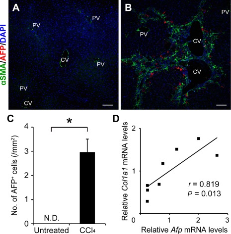
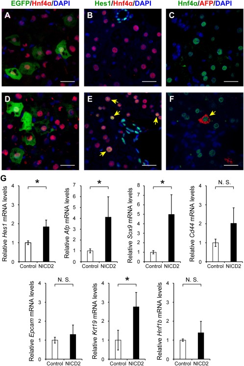
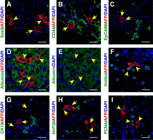
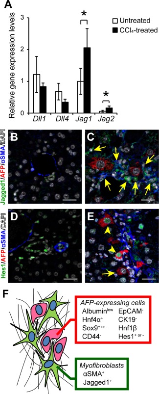
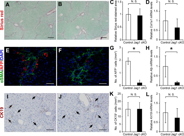
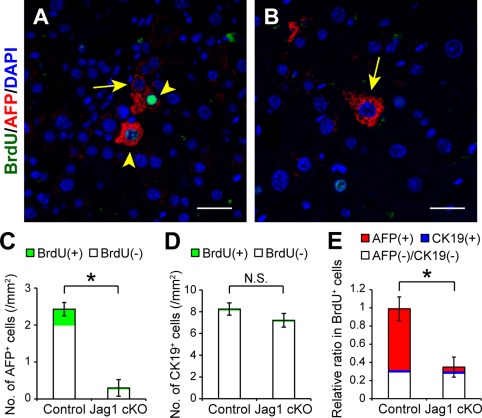
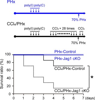
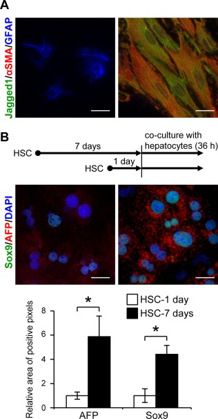
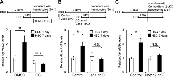
The liver is well known to possess high regenerative capacity in response to partial resection or tissue injury. However, liver regeneration is often impaired in the case of advanced liver fibrosis/cirrhosis when mature hepatocytes can hardly self-proliferate. Hepatic progenitor cells have been implicated as a source of hepatocytes in regeneration of the fibrotic liver. Although alpha-fetoprotein (AFP) is known as a clinical marker of progenitor cell induction in injured/fibrotic adult liver, the origin and features of such AFP-producing cells are not fully understood. Here, we demonstrate a unique and distinct AFP-expressing cell population that is induced by the Jagged1/Notch2 signal in murine fibrotic liver. Following repeated carbon tetrachloride injections, a significant number of AFP-positive cells with high proliferative ability were observed along the fibrous septa depending on the extent of liver fibrosis. These AFP-positive cells exhibited features of immature hepatocytes that were stained positively for hepatocyte-lineage markers, such as albumin and hepatocyte nuclear factor 4 alpha, and a stem/progenitor cell marker Sox9. A combination of immunohistological examination of fibrotic liver tissues and coculture experiments with primary hepatocytes and hepatic stellate cells indicated that increased Jagged1 expression in activated hepatic stellate cells stimulated Notch2 signaling and up-regulated AFP expression in adjacent hepatocytes. The mobilization and proliferation of AFP-positive cells in fibrotic liver were further enhanced after partial hepatectomy, which was significantly suppressed in Jagged1-conditional knockout mice. Finally, forced expression of the intracellular domain of Notch2 in normal liver induced a small number of AFP-expressing hepatocytes : Insight is provided into a novel pathophysiological role of Jagged1/Notch2 signaling in the induction of AFP-positive cells in fibrotic liver through the interaction between hepatocytes and activated hepatic stellate cells. ( 2017;1:215-229).

摘要

众所周知,肝脏在部分切除或组织损伤后具有很高的再生能力。然而,在晚期肝纤维化/肝硬化的情况下,当成熟肝细胞几乎无法自我增殖时,肝脏再生通常会受到损害。肝祖细胞被认为是纤维化肝脏再生中肝细胞的来源。尽管甲胎蛋白(AFP)是受损/纤维化成人肝脏中祖细胞诱导的临床标志物,但此类产生AFP的细胞的起源和特征尚未完全了解。在此,我们展示了在小鼠纤维化肝脏中由Jagged1/Notch2信号诱导产生的独特且不同的AFP表达细胞群。在反复注射四氯化碳后,根据肝纤维化程度,在纤维间隔中观察到大量具有高增殖能力的AFP阳性细胞。这些AFP阳性细胞表现出未成熟肝细胞的特征,对肝细胞谱系标志物如白蛋白和肝细胞核因子4α以及干细胞/祖细胞标志物Sox9呈阳性染色。纤维化肝组织的免疫组织学检查以及与原代肝细胞和肝星状细胞的共培养实验表明,活化肝星状细胞中Jagged1表达的增加刺激了Notch2信号传导,并上调了相邻肝细胞中AFP的表达。部分肝切除术后,纤维化肝脏中AFP阳性细胞的动员和增殖进一步增强,这在Jagged1条件性敲除小鼠中受到显著抑制。最后,在正常肝脏中强制表达Notch2的细胞内结构域诱导了少量表达AFP的肝细胞:通过肝细胞与活化肝星状细胞之间的相互作用,深入了解了Jagged1/Notch2信号传导在纤维化肝脏中诱导AFP阳性细胞的新病理生理作用。(2017;1:215 - 229)

https://cdn.ncbi.nlm.nih.gov/pmc/blobs/f6c8/5721449/46bf3e67a145/HEP4-1-215-g009.jpg
https://cdn.ncbi.nlm.nih.gov/pmc/blobs/f6c8/5721449/bf3b4a37f839/HEP4-1-215-g001.jpg
https://cdn.ncbi.nlm.nih.gov/pmc/blobs/f6c8/5721449/f34489ebf810/HEP4-1-215-g002.jpg
https://cdn.ncbi.nlm.nih.gov/pmc/blobs/f6c8/5721449/66ac0f9ab898/HEP4-1-215-g003.jpg
https://cdn.ncbi.nlm.nih.gov/pmc/blobs/f6c8/5721449/7e8f33865319/HEP4-1-215-g004.jpg
https://cdn.ncbi.nlm.nih.gov/pmc/blobs/f6c8/5721449/1e6f32de4873/HEP4-1-215-g005.jpg
https://cdn.ncbi.nlm.nih.gov/pmc/blobs/f6c8/5721449/1ce81e4b2787/HEP4-1-215-g006.jpg
https://cdn.ncbi.nlm.nih.gov/pmc/blobs/f6c8/5721449/636a84ea2ef2/HEP4-1-215-g007.jpg
https://cdn.ncbi.nlm.nih.gov/pmc/blobs/f6c8/5721449/12daa74c8f34/HEP4-1-215-g008.jpg
https://cdn.ncbi.nlm.nih.gov/pmc/blobs/f6c8/5721449/46bf3e67a145/HEP4-1-215-g009.jpg

相似文献

1
Identification of a novel alpha-fetoprotein-expressing cell population induced by the Jagged1/Notch2 signal in murine fibrotic liver.在小鼠纤维化肝脏中由Jagged1/Notch2信号诱导的新型甲胎蛋白表达细胞群的鉴定
Hepatol Commun. 2017 Mar 27;1(3):215-229. doi: 10.1002/hep4.1026. eCollection 2017 May.
2
Cooperation of liver cells in health and disease.健康与疾病状态下肝细胞的协作。
Adv Anat Embryol Cell Biol. 2001;161:III-XIII, 1-151. doi: 10.1007/978-3-642-56553-3.
3
Hepatic progenitor cells, stem cells, and AFP expression in models of liver injury.肝损伤模型中的肝祖细胞、干细胞及甲胎蛋白表达
Int J Exp Pathol. 2006 Oct;87(5):343-59. doi: 10.1111/j.1365-2613.2006.00485.x.
4
Identification of a Novel Bone Marrow Cell-Derived Accelerator of Fibrotic Liver Regeneration Through Mobilization of Hepatic Progenitor Cells in Mice.通过动员小鼠肝祖细胞鉴定新型骨髓细胞来源的肝纤维化再生促进剂。
Stem Cells. 2019 Jan;37(1):89-101. doi: 10.1002/stem.2916. Epub 2018 Oct 31.
5
Hepatic stellate cells regulate hepatic progenitor cells differentiation via the TGF-β1/Jagged1 signaling axis.肝星状细胞通过 TGF-β1/Jagged1 信号轴调节肝祖细胞分化。
J Cell Physiol. 2019 Jun;234(6):9283-9296. doi: 10.1002/jcp.27609. Epub 2018 Oct 14.
6
TGF-β1 signaling regulates mouse hepatic stellate cell differentiation via the Jagged1/Notch pathway.转化生长因子-β1信号通路通过Jagged1/Notch途径调控小鼠肝星状细胞分化。
Life Sci. 2018 Jan 1;192:221-230. doi: 10.1016/j.lfs.2017.11.018. Epub 2017 Nov 14.
7
Activation, proliferation, and differentiation of progenitor cells into hepatocytes in the D-galactosamine model of liver regeneration.在D-半乳糖胺肝再生模型中祖细胞向肝细胞的激活、增殖和分化。
Am J Pathol. 1993 Dec;143(6):1606-20.
8
Expansion and Hepatic Differentiation of Adult Blood-Derived CD34+ Progenitor Cells and Promotion of Liver Regeneration After Acute Injury.成年血液来源的CD34+祖细胞的扩增与肝分化以及急性损伤后肝脏再生的促进
Stem Cells Transl Med. 2016 Jun;5(6):723-32. doi: 10.5966/sctm.2015-0268. Epub 2016 Apr 13.
9
In vitro differentiated hepatic oval-like cells enhance hepatic regeneration in CCl -induced hepatic injury.体外分化的肝卵圆样细胞增强了四氯化碳诱导的肝损伤中的肝再生。
Cell Biol Int. 2017 Jan;41(1):51-61. doi: 10.1002/cbin.10699. Epub 2016 Nov 13.
10
Involvement of the nuclear high mobility group B1 peptides released from injured hepatocytes in murine hepatic fibrogenesis.受损肝细胞释放的核高迁移率族蛋白B1肽参与小鼠肝纤维化形成过程。
Biochim Biophys Acta. 2014 Sep;1842(9):1720-32. doi: 10.1016/j.bbadis.2014.06.017. Epub 2014 Jun 23.

引用本文的文献

1
Communication initiated by hepatocytes: The driver of HSC activation and liver fibrosis.肝细胞引发的通讯:肝星状细胞激活和肝纤维化的驱动因素
Hepatol Commun. 2025 Jul 14;9(8). doi: 10.1097/HC9.0000000000000753. eCollection 2025 Aug 1.
2
Pay attention to the value of liver regeneration in the re-compensation of decompensated cirrhosis.关注肝再生在失代偿期肝硬化再代偿中的价值。
World J Gastroenterol. 2025 Jun 28;31(24):106564. doi: 10.3748/wjg.v31.i24.106564.
3
Jag1 insufficiency alters liver fibrosis via T cell and hepatocyte differentiation defects.

本文引用的文献

1
Conversion of Terminally Committed Hepatocytes to Culturable Bipotent Progenitor Cells with Regenerative Capacity.终末分化的肝细胞向具有再生能力的可培养双能祖细胞的转化。
Cell Stem Cell. 2017 Jan 5;20(1):41-55. doi: 10.1016/j.stem.2016.10.007. Epub 2016 Nov 10.
2
Kupffer cells induce Notch-mediated hepatocyte conversion in a common mouse model of intrahepatic cholangiocarcinoma.枯否细胞在肝内胆管癌的一种常见小鼠模型中诱导 Notch 介导的肝细胞转化。
Sci Rep. 2016 Oct 4;6:34691. doi: 10.1038/srep34691.
3
Foetal hepatic progenitor cells assume a cholangiocytic cell phenotype during two-dimensional pre-culture.
Jag1 不足通过 T 细胞和肝细胞分化缺陷改变肝纤维化。
EMBO Mol Med. 2024 Nov;16(11):2946-2975. doi: 10.1038/s44321-024-00145-8. Epub 2024 Oct 2.
4
Saffron reduces the liver fibrosis in mice by inhibiting the JAK/STAT3 pathway.藏红花通过抑制 JAK/STAT3 通路减少小鼠肝纤维化。
Acta Cir Bras. 2023 Dec 1;38:e385823. doi: 10.1590/acb385823. eCollection 2023.
5
Cellular heterogeneity and plasticity during NAFLD progression.非酒精性脂肪性肝病进展过程中的细胞异质性和可塑性。
Front Mol Biosci. 2023 Aug 11;10:1221669. doi: 10.3389/fmolb.2023.1221669. eCollection 2023.
6
Serum Alpha-Fetoprotein as a Predictor of Liver Fibrosis in HBeAg-Positive Chronic Hepatitis B Patients.血清甲胎蛋白作为 HBeAg 阳性慢性乙型肝炎患者肝纤维化的预测指标。
Medicina (Kaunas). 2023 May 11;59(5):923. doi: 10.3390/medicina59050923.
7
Mitochondrial dysfunction reactivates α-fetoprotein expression that drives copper-dependent immunosuppression in mitochondrial disease models.线粒体功能障碍重新激活甲胎蛋白表达,从而在线粒体疾病模型中驱动铜依赖性免疫抑制。
J Clin Invest. 2023 Jan 3;133(1):e154684. doi: 10.1172/JCI154684.
8
In vivo partial cellular reprogramming enhances liver plasticity and regeneration.体内部分细胞重编程增强肝脏的可塑性和再生能力。
Cell Rep. 2022 Apr 26;39(4):110730. doi: 10.1016/j.celrep.2022.110730.
9
Therapeutic potential of macrophage colony-stimulating factor in chronic liver disease.巨噬细胞集落刺激因子在慢性肝病中的治疗潜力。
Dis Model Mech. 2022 Apr 1;15(4). doi: 10.1242/dmm.049387. Epub 2022 Apr 19.
10
Two distinct Notch signals, Delta-like 4/Notch1 and Jagged-1/Notch2, antagonistically regulate chemical hepatocarcinogenesis in mice.两种不同的 Notch 信号,Delta-like 4/Notch1 和 Jagged-1/Notch2,在小鼠中拮抗调节化学性肝癌发生。
Commun Biol. 2022 Jan 21;5(1):85. doi: 10.1038/s42003-022-03013-8.
在二维原代培养过程中,胎儿肝祖细胞呈现出胆管细胞表型。
Sci Rep. 2016 Jun 23;6:28283. doi: 10.1038/srep28283.
4
Intrahepatic bile ducts are developed through formation of homogeneous continuous luminal network and its dynamic rearrangement in mice.肝内胆管通过在小鼠体内形成均质连续的腔道网络及其动态重排而发育。
Hepatology. 2016 Jul;64(1):175-88. doi: 10.1002/hep.28521. Epub 2016 Apr 4.
5
Stem cells versus plasticity in liver and pancreas regeneration.肝和胰腺再生中的干细胞与可塑性。
Nat Cell Biol. 2016 Mar;18(3):238-45. doi: 10.1038/ncb3309.
6
A Combination of Mitochondrial Oxidative Stress and Excess Fat/Calorie Intake Accelerates Steatohepatitis by Enhancing Hepatic CC Chemokine Production in Mice.线粒体氧化应激与脂肪/热量摄入过多相结合,通过增强小鼠肝脏CC趋化因子的产生加速脂肪性肝炎。
PLoS One. 2016 Jan 8;11(1):e0146592. doi: 10.1371/journal.pone.0146592. eCollection 2016.
7
Disappearance of centroacinar cells in the Notch ligand-deficient pancreas.Notch配体缺陷型胰腺中腺泡中心细胞的消失。
Genes Cells. 2015 Jun;20(6):500-11. doi: 10.1111/gtc.12243. Epub 2015 Apr 27.
8
Impact of Sox9 dosage and Hes1-mediated Notch signaling in controlling the plasticity of adult pancreatic duct cells in mice.Sox9剂量和Hes1介导的Notch信号对小鼠成年胰腺导管细胞可塑性的影响
Sci Rep. 2015 Feb 17;5:8518. doi: 10.1038/srep08518.
9
Alpha-fetoprotein: A biomarker for the recruitment of progenitor cells in the liver in patients with acute liver injury or failure.甲胎蛋白:急性肝损伤或肝衰竭患者肝脏中祖细胞募集的生物标志物。
Hepatol Res. 2015 Oct;45(10):E12-20. doi: 10.1111/hepr.12448. Epub 2014 Dec 19.
10
Bipotential adult liver progenitors are derived from chronically injured mature hepatocytes.双潜能成年肝脏祖细胞源自长期受损的成熟肝细胞。
Cell Stem Cell. 2014 Nov 6;15(5):605-18. doi: 10.1016/j.stem.2014.09.008. Epub 2014 Oct 9.