Department of Pharmaceutical Sciences, University of Piemonte Orientale "A. Avogadro", Novara, Italy.
NIS Interdepartmental Center, University of Torino, Torino, Italy.
Sci Rep. 2018 Feb 9;8(1):2760. doi: 10.1038/s41598-018-21157-8.
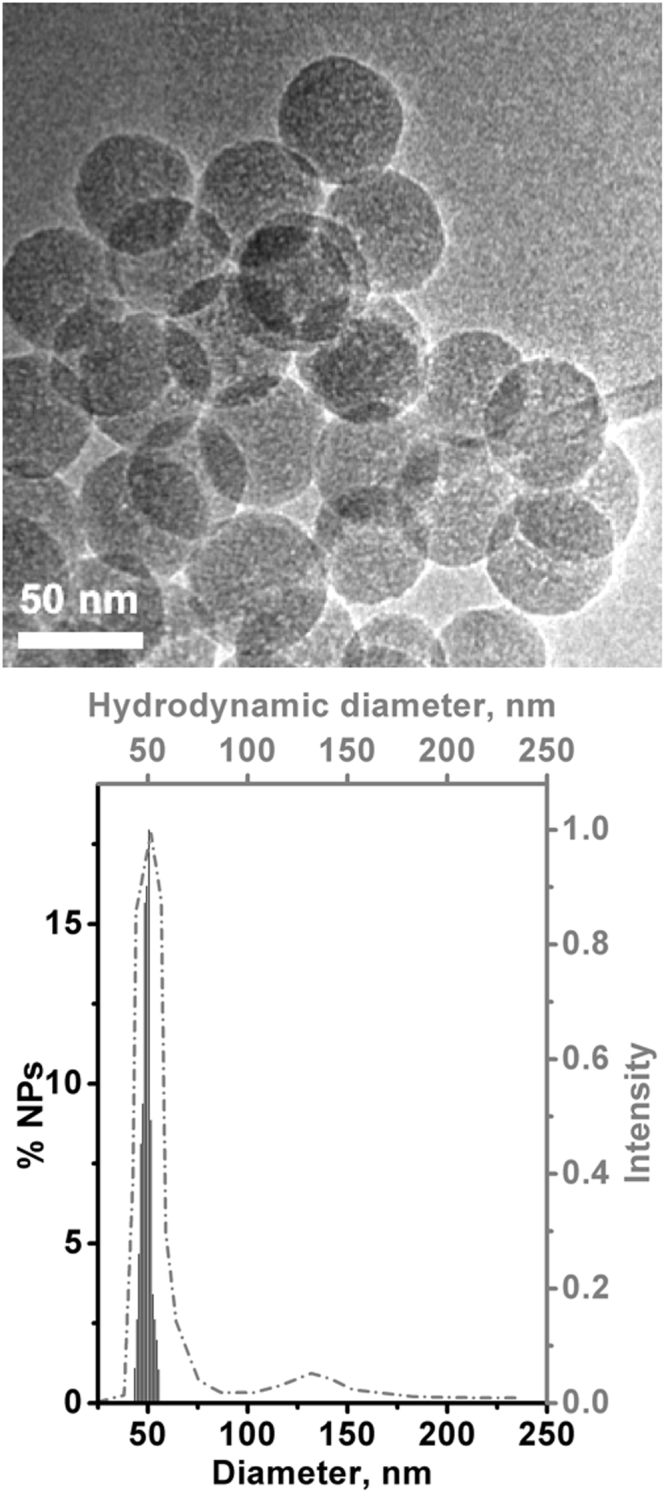
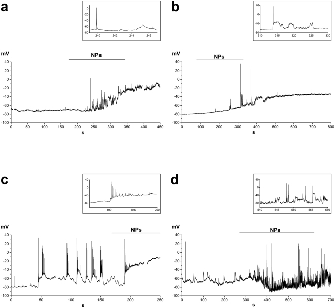
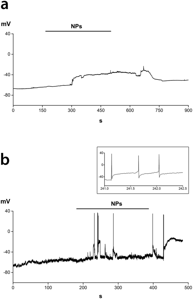
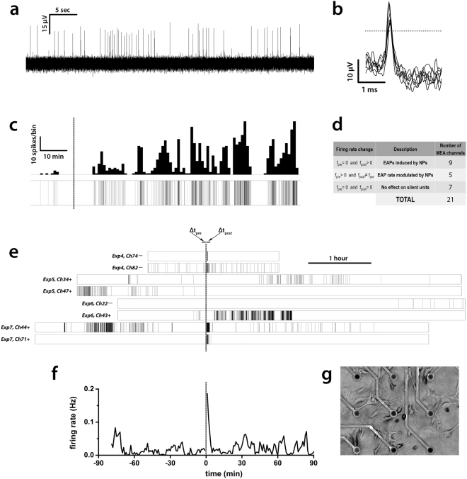
Engineered silica nanoparticles (NPs) have attracted increasing interest in several applications, and particularly in the field of nanomedicine, thanks to the high biocompatibility of this material. For their optimal and controlled use, the understanding of the mechanisms elicited by their interaction with the biological target is a prerequisite, especially when dealing with cells particularly vulnerable to environmental stimuli like neurons. Here we have combined different electrophysiological approaches (both at the single cell and at the population level) with a genomic screening in order to analyze, in GT1-7 neuroendocrine cells, the impact of SiO NPs (50 ± 3 nm in diameter) on electrical activity and gene expression, providing a detailed analysis of the impact of a nanoparticle on neuronal excitability. We find that 20 µg mL NPs induce depolarization of the membrane potential, with a modulation of the firing of action potentials. Recordings of electrical activity with multielectrode arrays provide further evidence that the NPs evoke a temporary increase in firing frequency, without affecting the functional behavior on a time scale of hours. Finally, NPs incubation up to 24 hours does not induce any change in gene expression.
经过工程设计的硅纳米颗粒(NPs)由于其材料具有高生物兼容性,在多个应用领域中,特别是在纳米医学领域,引起了越来越多的关注。为了实现其最佳和可控的使用,理解其与生物靶标相互作用所引发的机制是先决条件,特别是在处理对环境刺激(如神经元)特别敏感的细胞时。在这里,我们结合了不同的电生理方法(单细胞和群体水平)以及基因组筛选,以便在 GT1-7 神经内分泌细胞中分析 SiO2 NPs(直径为 50±3nm)对电活动和基因表达的影响,对纳米颗粒对神经元兴奋性的影响进行了详细的分析。我们发现,20μg/mL 的 NPs 会引起膜电位去极化,动作电位的发放也会随之调制。通过多电极阵列进行电活动记录进一步证明,NPs 会引起短暂的发放频率增加,而不会在数小时的时间尺度上影响其功能行为。最后,NPs 孵育 24 小时不会引起基因表达的任何变化。