Laboratory of Neurodegenerative Diseases, School of Biomedical Sciences, LKS Faculty of Medicine, The University of Hong Kong, Pokfulam, Hong Kong, SAR, China.
Present address: Nanjing Key Laboratory of Pediatrics, Children's Hospital of Nanjing Medical University, Nanjing, 210008, China.
Part Fibre Toxicol. 2018 Jul 3;15(1):28. doi: 10.1186/s12989-018-0263-3.
Silica nanoparticles (SiO-NPs) are naturally enriched and broadly utilized in the manufacturing industry. While previous studies have demonstrated toxicity in neuronal cell lines after SiO-NPs exposure, the role of SiO-NPs in neurodegeneration is largely unknown. Here, we evaluated the effects of SiO-NPs-exposure on behavior, neuropathology, and synapse in young adult mice and primary cortical neuron cultures.
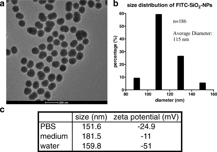
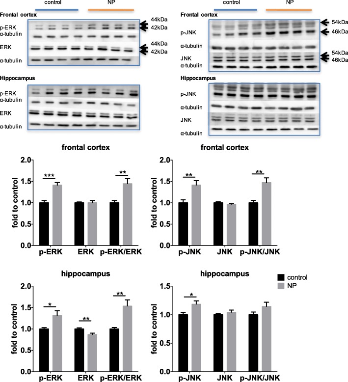
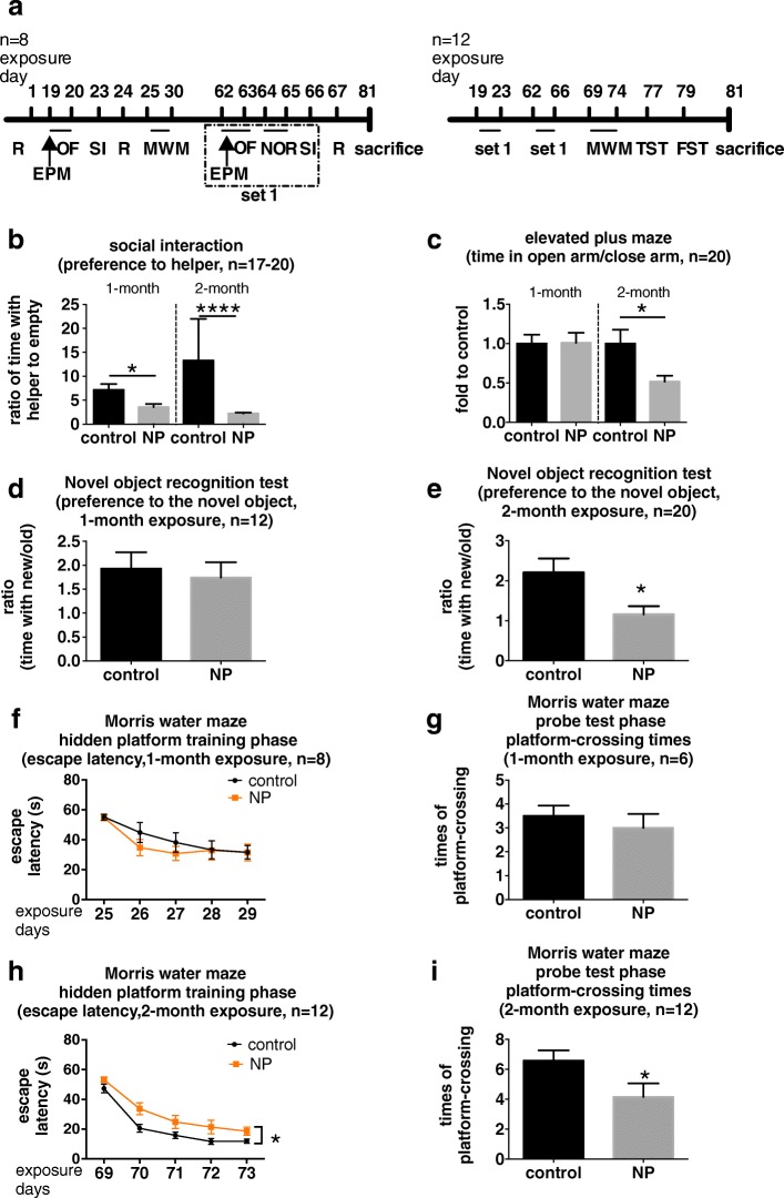
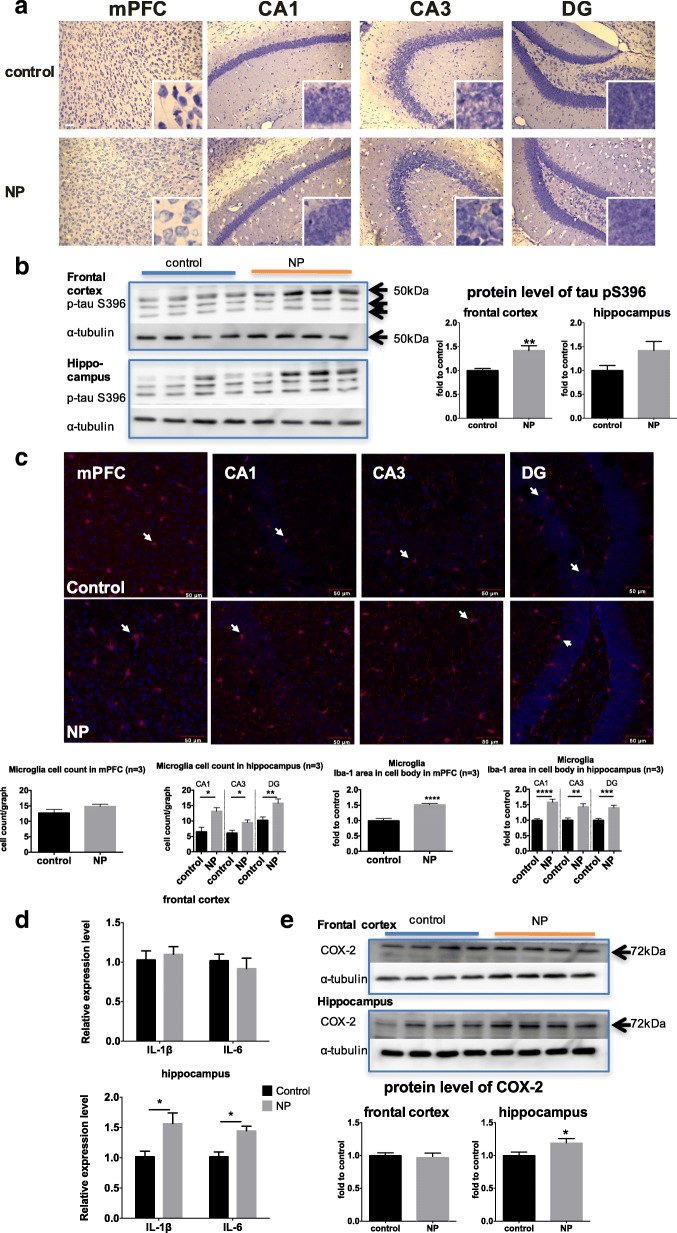
Male C57BL/6 N mice (3 months old) were exposed to either vehicle (sterile PBS) or fluorescein isothiocyanate (FITC)-tagged SiO-NPs (NP) using intranasal instillation. Behavioral tests were performed after 1 and 2 months of exposure. We observed decreased social activity at both time points as well as anxiety and cognitive impairment after 2 months in the NP-exposed mice. NP deposition was primarily detected in the medial prefrontal cortex and the hippocampus. Neurodegeneration-like pathological changes, including reduced Nissl staining, increased tau phosphorylation, and neuroinflammation, were also present in the brains of NP-exposed mice. Furthermore, we observed NP-induced impairment in exocytosis along with decreased synapsin I and increased synaptophysin expression in the synaptosome fractions isolated from the frontal cortex as well as primary neuronal cultures. Extracellular signal-regulated kinase (ERK) and c-Jun N-terminal kinase (JNK) were also activated in the frontal cortex of NP-exposed mice. Moreover, inhibition of ERK activation prevented NP-mediated changes in exocytosis in cultured neurons, highlighting a key role in the changes induced by NP exposure.
Intranasal instillation of SiO-NPs results in mood dysfunction and cognitive impairment in young adult mice and causes neurodegeneration-like pathology and synaptic changes via ERK activation.
硅纳米颗粒(SiO-NPs)在制造业中自然富集并广泛应用。虽然之前的研究表明,SiO-NPs 暴露后神经元细胞系存在毒性,但 SiO-NPs 在神经退行性变中的作用还知之甚少。在这里,我们评估了 SiO-NPs 暴露对年轻成年小鼠和原代皮质神经元培养物的行为、神经病理学和突触的影响。
雄性 C57BL/6N 小鼠(3 个月大)通过鼻腔内滴注接受载体(无菌 PBS)或异硫氰酸荧光素(FITC)标记的 SiO-NPs(NP)。暴露后 1 个月和 2 个月进行行为测试。我们观察到,NP 暴露的小鼠在这两个时间点的社交活动减少,以及在 2 个月后的焦虑和认知障碍。NP 沉积主要在中前额皮质和海马体中检测到。NP 暴露的小鼠大脑中还存在神经退行性样病理变化,包括尼氏染色减少、tau 磷酸化增加和神经炎症。此外,我们观察到 NP 诱导的突触小体从质膜出泡作用受损,以及从额皮质分离的突触小体和原代神经元培养物中突触素 I 减少和突触小体蛋白表达增加。NP 暴露的小鼠前额皮质中细胞外信号调节激酶(ERK)和 c-Jun N 端激酶(JNK)也被激活。此外,ERK 激活的抑制可防止 NP 介导的培养神经元中出泡作用的改变,突出了 NP 暴露引起的改变的关键作用。
SiO-NPs 的鼻腔内滴注导致年轻成年小鼠出现情绪功能障碍和认知障碍,并通过 ERK 激活导致神经退行性样病理和突触改变。