School of Engineering, University of Glasgow, Glasgow, United Kingdom.
Department of Engineering Science, University of Oxford, Oxford, United Kingdom.
Appl Environ Microbiol. 2018 Apr 2;84(8). doi: 10.1128/AEM.02508-17. Print 2018 Apr 15.
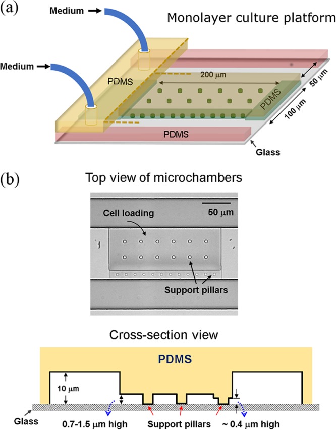
Lasers are instrumental in advanced bioimaging and Raman spectroscopy. However, they are also well known for their destructive effects on living organisms, leading to concerns about the adverse effects of laser technologies. To implement Raman spectroscopy for cell analysis and manipulation, such as Raman-activated cell sorting, it is crucial to identify nondestructive conditions for living cells. Here, we evaluated quantitatively the effect of 532-nm laser irradiation on bacterial cell fate and growth at the single-cell level. Using a purpose-built microfluidic platform, we were able to quantify the growth characteristics, i.e., specific growth rates and lag times of individual cells, as well as the survival rate of a population in conjunction with Raman spectroscopy. Representative Gram-negative and Gram-positive species show similar trends in response to a laser irradiation dose. Laser irradiation could compromise the physiological function of cells, and the degree of destruction is both dose and strain dependent, ranging from reduced cell growth to a complete loss of cell metabolic activity and finally to physical disintegration. Gram-positive bacterial cells are more susceptible than Gram-negative bacterial strains to irradiation-induced damage. By directly correlating Raman acquisition with single-cell growth characteristics, we provide evidence of nondestructive characteristics of Raman spectroscopy on individual bacterial cells. However, while strong Raman signals can be obtained without causing cell death, the variety of responses from different strains and from individual cells justifies careful evaluation of Raman acquisition conditions if cell viability is critical. In Raman spectroscopy, the use of powerful monochromatic light in laser-based systems facilitates the detection of inherently weak signals. This allows environmentally and clinically relevant microorganisms to be measured at the single-cell level. The significance of being able to perform Raman measurement is that, unlike label-based fluorescence techniques, it provides a "fingerprint" that is specific to the identity and state of any (unlabeled) sample. Thus, it has emerged as a powerful method for studying living cells under physiological and environmental conditions. However, the laser's high power also has the potential to kill bacteria, which leads to concerns. The research presented here is a quantitative evaluation that provides a generic platform and methodology to evaluate the effects of laser irradiation on individual bacterial cells. Furthermore, it illustrates this by determining the conditions required to nondestructively measure the spectra of representative bacteria from several different groups.
激光在先进的生物成像和拉曼光谱学中起着重要作用。然而,它们也因对生物体的破坏性影响而广为人知,这引发了人们对激光技术的不良影响的关注。为了在细胞分析和操作中实施拉曼光谱学,例如拉曼激活细胞分选,确定对活细胞无损伤的条件至关重要。在这里,我们在单细胞水平上定量评估了 532nm 激光照射对细菌细胞命运和生长的影响。使用专门构建的微流控平台,我们能够定量测量单个细胞的生长特性,即特定生长速率和滞后时间,以及群体的存活率,并结合拉曼光谱进行分析。有代表性的革兰氏阴性和革兰氏阳性菌对激光辐照剂量表现出相似的响应趋势。激光照射可能会损害细胞的生理功能,破坏程度既依赖于剂量又依赖于菌株,范围从细胞生长减少到细胞代谢活性完全丧失,最后到物理解体。革兰氏阳性细菌细胞比革兰氏阴性细菌菌株更容易受到辐照损伤。通过将拉曼采集与单细胞生长特性直接相关联,我们提供了拉曼光谱对单个细菌细胞无损伤特性的证据。然而,虽然可以在不导致细胞死亡的情况下获得强拉曼信号,但不同菌株和单个细胞的各种响应表明,如果细胞活力至关重要,则需要仔细评估拉曼采集条件。在拉曼光谱学中,基于激光的系统中使用强大的单色光有助于检测固有较弱的信号。这使得可以在单细胞水平上测量环境和临床相关的微生物。能够进行拉曼测量的意义在于,与基于标记的荧光技术不同,它提供了与任何(未标记)样本的身份和状态相关的“指纹”。因此,它已成为在生理和环境条件下研究活细胞的强大方法。然而,激光的高功率也有可能杀死细菌,这引起了人们的关注。本研究是对激光照射对单个细菌细胞影响的定量评估,提供了一个通用的平台和方法来评估激光照射对单个细菌细胞的影响。此外,它通过确定从几个不同群体中代表细菌的非破坏性测量光谱所需的条件来说明这一点。