Seo NaRi, Lee Sung-Ho, Ju Kyung Won, Woo JaeMan, Kim BongJu, Kim SoungMin, Jahng Jeong Won, Lee Jong-Ho
Department of Oral and Maxillofacial Surgery, Graduate School of Dentistry, Seoul National University; Dental Research Institute, Seoul National University, Seoul, South Korea.
Department of Oral and Maxillofacial Surgery, Seoul National University Dental Hospital; Dental Research Institute, Seoul National University, Seoul, South Korea.
Neural Regen Res. 2018 Jan;13(1):145-153. doi: 10.4103/1673-5374.224383.
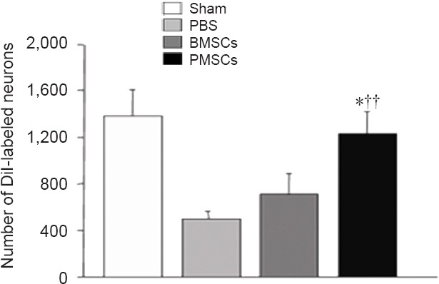
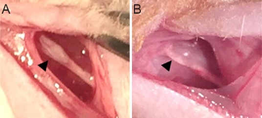
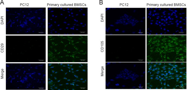
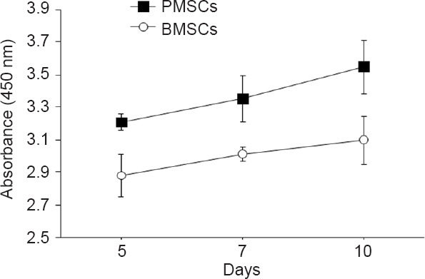
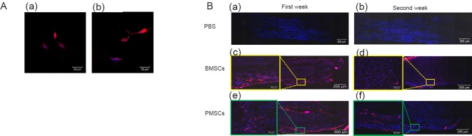
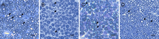
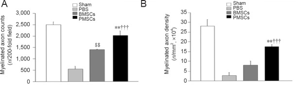
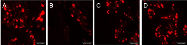
Bone marrow-derived mesenchymal stem cells (BMSCs) have been shown to promote the regeneration of injured peripheral nerves. Pulsed electromagnetic field (PEMF) reportedly promotes the proliferation and neuronal differentiation of BMSCs. Low-frequency PEMF can induce the neuronal differentiation of BMSCs in the absence of nerve growth factors. This study was designed to investigate the effects of low-frequency PEMF pretreatment on the proliferation and function of BMSCs and the effects of low-frequency PEMF pre-treated BMSCs on the regeneration of injured peripheral nerve using in vitro and in vivo experiments. In in vitro experiments, quantitative DNA analysis was performed to determine the proliferation of BMSCs, and reverse transcription-polymerase chain reaction was performed to detect S100 (Schwann cell marker), glial fibrillary acidic protein (astrocyte marker), and brain-derived neurotrophic factor and nerve growth factor (neurotrophic factors) mRNA expression. In the in vivo experiments, rat models of crush-injured mental nerve established using clamp method were randomly injected with low-frequency PEMF pretreated BMSCs, unpretreated BMSCs or PBS at the injury site (1 × 10 cells). DiI-labeled BMSCs injected at the injury site were counted under the fluorescence microscope to determine cell survival. One or two weeks after cell injection, functional recovery of the injured nerve was assessed using the sensory test with von Frey filaments. Two weeks after cell injection, axonal regeneration was evaluated using histomorphometric analysis and retrograde labeling of trigeminal ganglion neurons. In vitro experiment results revealed that low-frequency PEMF pretreated BMSCs proliferated faster and had greater mRNA expression of growth factors than unpretreated BMSCs. In vivo experiment results revealed that compared with injection of unpretreated BMSCs, injection of low-frequency PEMF pretreated BMSCs led to higher myelinated axon count and axon density and more DiI-labeled neurons in the trigeminal ganglia, contributing to rapider functional recovery of injured mental nerve. These findings suggest that low-frequency PEMF pretreatment is a promising approach to enhance the efficacy of cell therapy for peripheral nerve injury repair.
骨髓间充质干细胞(BMSCs)已被证明可促进受损周围神经的再生。据报道,脉冲电磁场(PEMF)可促进BMSCs的增殖和神经元分化。低频PEMF在无神经生长因子的情况下可诱导BMSCs的神经元分化。本研究旨在通过体外和体内实验,研究低频PEMF预处理对BMSCs增殖和功能的影响,以及低频PEMF预处理的BMSCs对受损周围神经再生的影响。在体外实验中,进行定量DNA分析以确定BMSCs的增殖情况,并进行逆转录-聚合酶链反应以检测S100(雪旺细胞标志物)、胶质纤维酸性蛋白(星形胶质细胞标志物)以及脑源性神经营养因子和神经生长因子(神经营养因子)的mRNA表达。在体内实验中,使用钳夹法建立大鼠颏神经挤压损伤模型,在损伤部位(1×10个细胞)随机注射低频PEMF预处理的BMSCs、未预处理的BMSCs或PBS。在荧光显微镜下计数注射到损伤部位的DiI标记的BMSCs,以确定细胞存活情况。细胞注射后1或2周,使用von Frey细丝进行感觉测试评估受损神经的功能恢复情况。细胞注射后2周,使用组织形态计量分析和三叉神经节神经元逆行标记评估轴突再生情况。体外实验结果显示,低频PEMF预处理的BMSCs比未预处理的BMSCs增殖更快,生长因子的mRNA表达更高。体内实验结果显示,与注射未预处理的BMSCs相比,注射低频PEMF预处理的BMSCs导致三叉神经节中有更高的有髓轴突计数和轴突密度以及更多的DiI标记神经元,有助于受损颏神经更快地功能恢复。这些发现表明,低频PEMF预处理是一种有前景的方法,可提高细胞疗法修复周围神经损伤的疗效。