• 文献检索
  • 文档翻译
  • 深度研究
  • 学术资讯
  • Suppr Zotero 插件Zotero 插件
  • 邀请有礼
  • 套餐&价格
  • 历史记录
应用&插件
Suppr Zotero 插件Zotero 插件浏览器插件Mac 客户端Windows 客户端微信小程序
定价
高级版会员购买积分包购买API积分包
服务
文献检索文档翻译深度研究API 文档MCP 服务
关于我们
关于 Suppr公司介绍联系我们用户协议隐私条款
关注我们

Suppr 超能文献

核心技术专利:CN118964589B侵权必究
粤ICP备2023148730 号-1Suppr @ 2026

文献检索

告别复杂PubMed语法,用中文像聊天一样搜索,搜遍4000万医学文献。AI智能推荐,让科研检索更轻松。

立即免费搜索

文件翻译

保留排版,准确专业,支持PDF/Word/PPT等文件格式,支持 12+语言互译。

免费翻译文档

深度研究

AI帮你快速写综述,25分钟生成高质量综述,智能提取关键信息,辅助科研写作。

立即免费体验

心血管磁共振全自动像素定量心肌灌注成像的诊断性能。

Diagnostic Performance of Fully Automated Pixel-Wise Quantitative Myocardial Perfusion Imaging by Cardiovascular Magnetic Resonance.

机构信息

National Heart, Lung, and Blood Institute, National Institutes of Health, Bethesda, Maryland.

National Heart, Lung, and Blood Institute, National Institutes of Health, Bethesda, Maryland.

出版信息

JACC Cardiovasc Imaging. 2018 May;11(5):697-707. doi: 10.1016/j.jcmg.2018.01.005. Epub 2018 Feb 14.

DOI:10.1016/j.jcmg.2018.01.005
PMID:29454767
原文链接:https://pmc.ncbi.nlm.nih.gov/articles/PMC8760891/
Abstract

OBJECTIVES

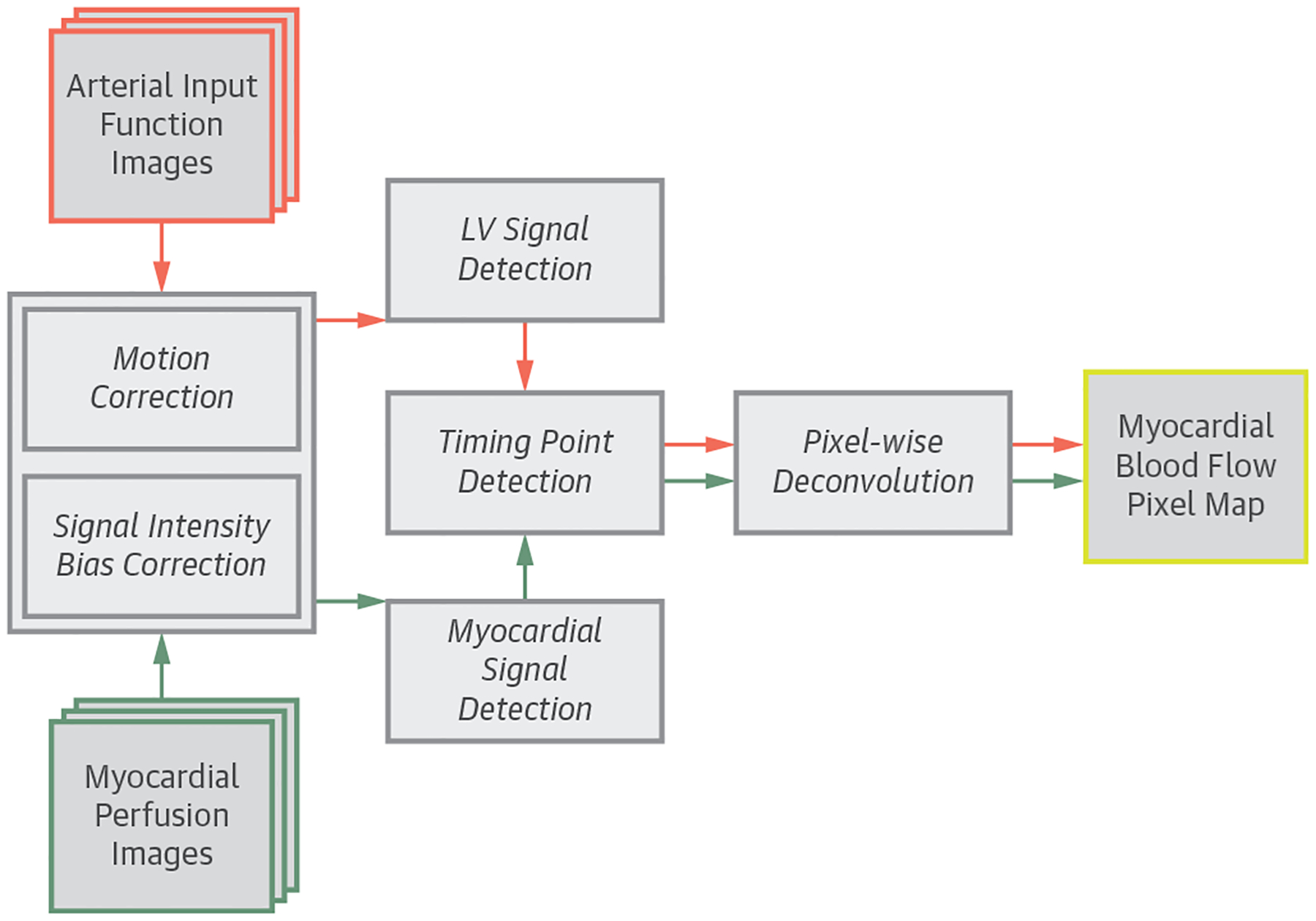
The authors developed a fully automated framework to quantify myocardial blood flow (MBF) from contrast-enhanced cardiac magnetic resonance (CMR) perfusion imaging and evaluated its diagnostic performance in patients.

BACKGROUND

Fully quantitative CMR perfusion pixel maps were previously validated with microsphere MBF measurements and showed potential in clinical applications, but the methods required laborious manual processes and were excessively time-consuming.

METHODS

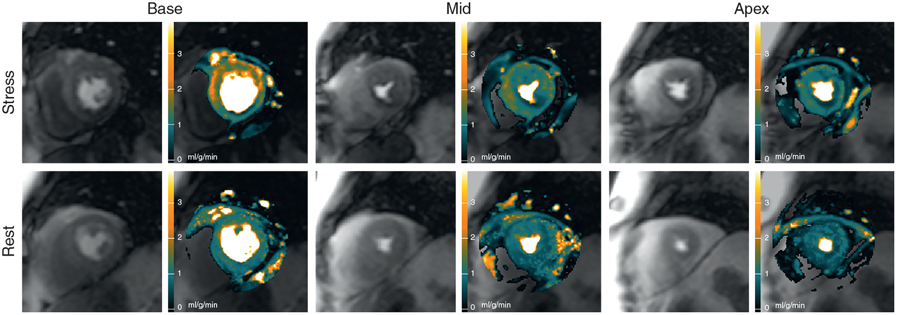
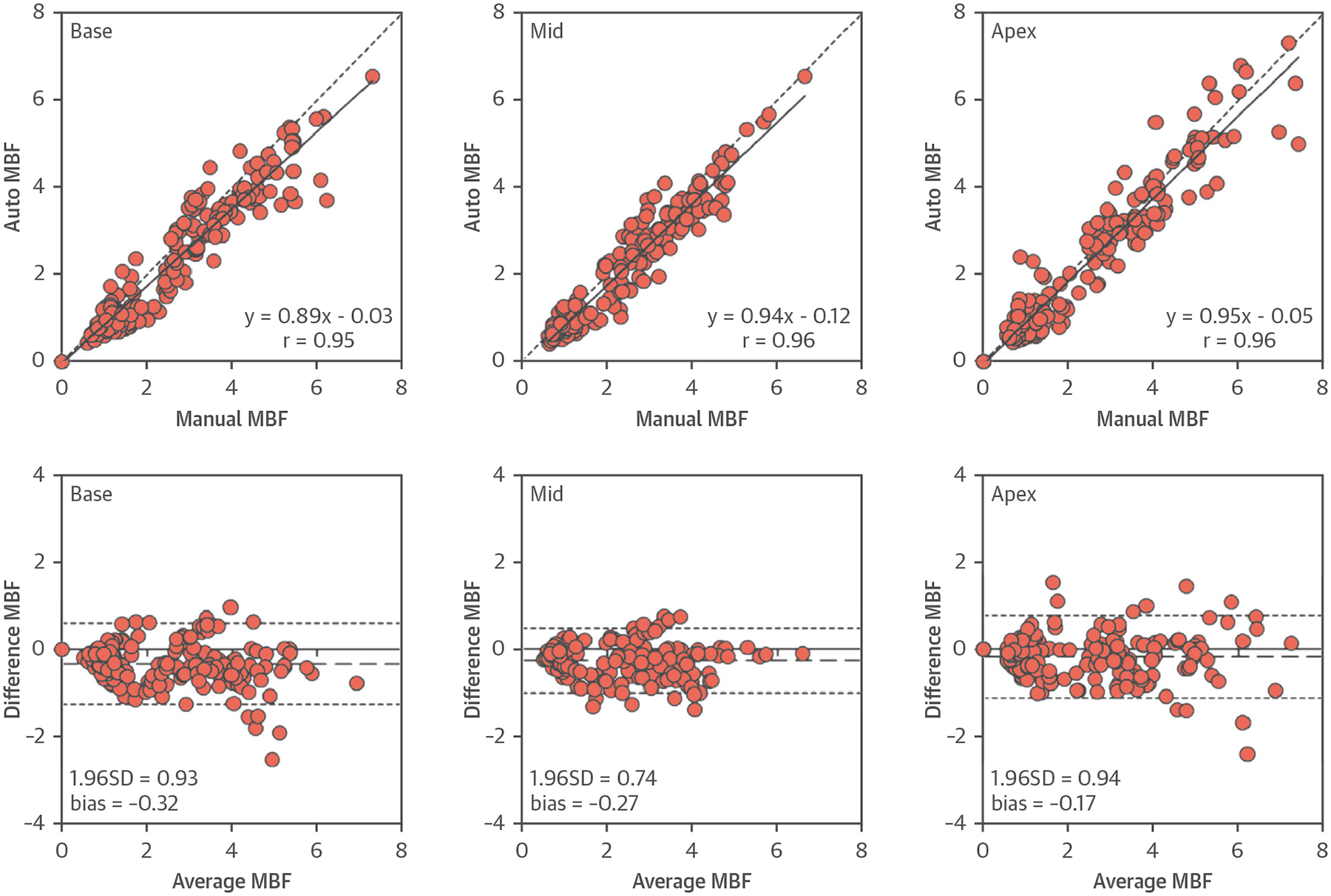
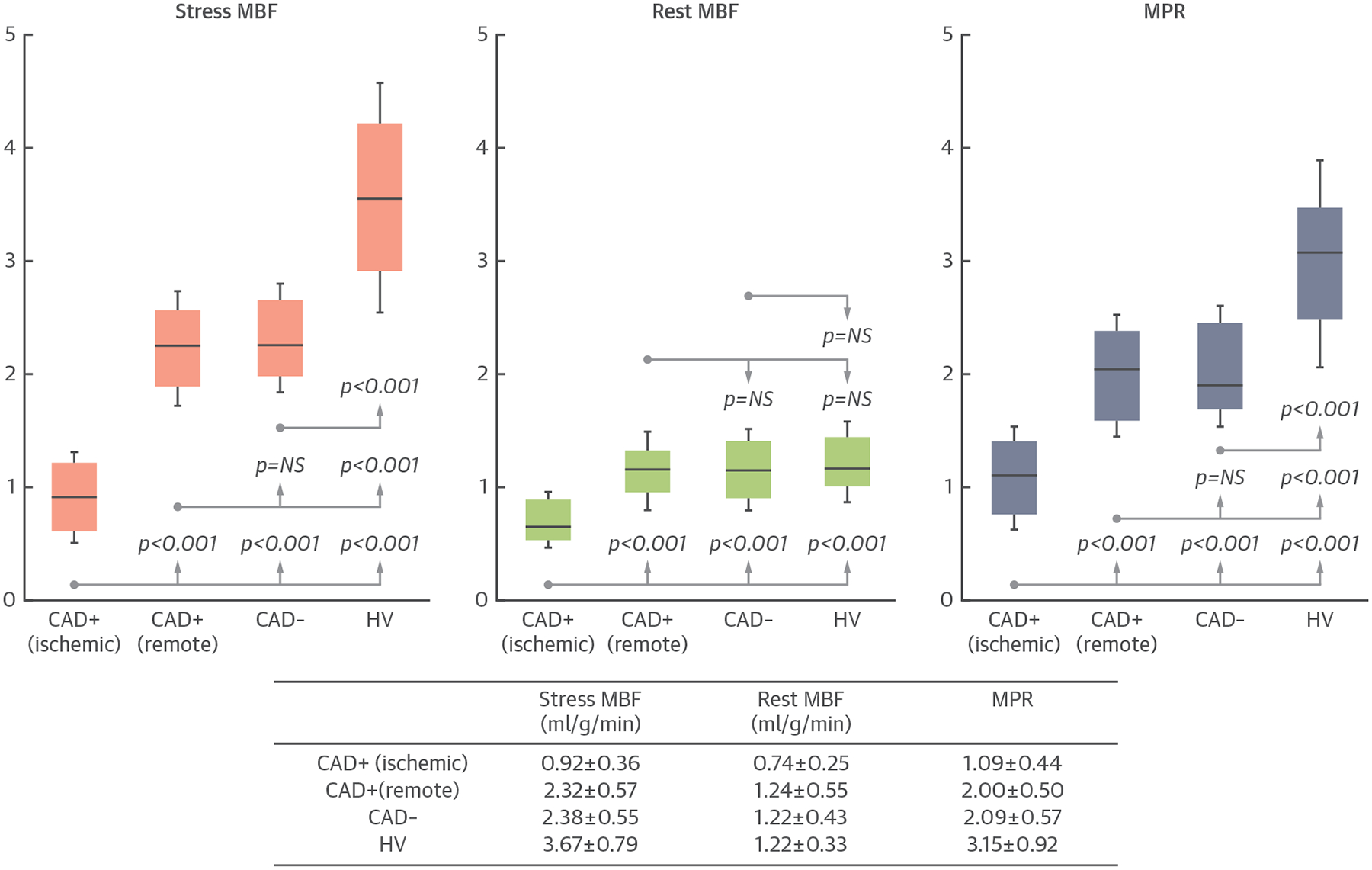
CMR perfusion imaging was performed on 80 patients with known or suspected coronary artery disease (CAD) and 17 healthy volunteers. Significant CAD was defined by quantitative coronary angiography (QCA) as ≥70% stenosis. Nonsignificant CAD was defined by: 1) QCA as <70% stenosis; or 2) coronary computed tomography angiography as <30% stenosis and a calcium score of 0 in all vessels. Automatically generated MBF maps were compared with manual quantification on healthy volunteers. Diagnostic performance of the automated MBF pixel maps was analyzed on patients using absolute MBF, myocardial perfusion reserve (MPR), and relative measurements of MBF and MPR.

RESULTS

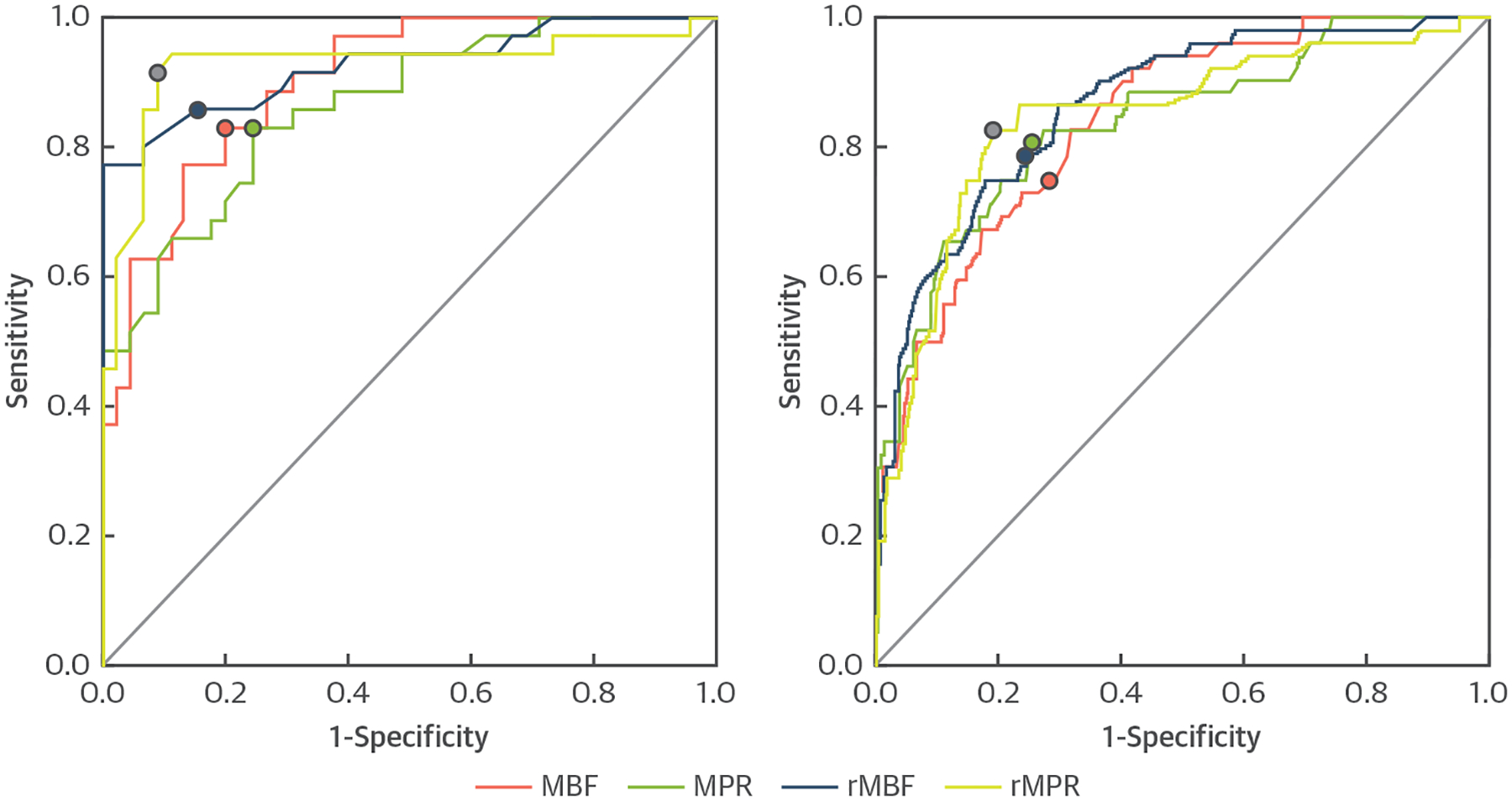
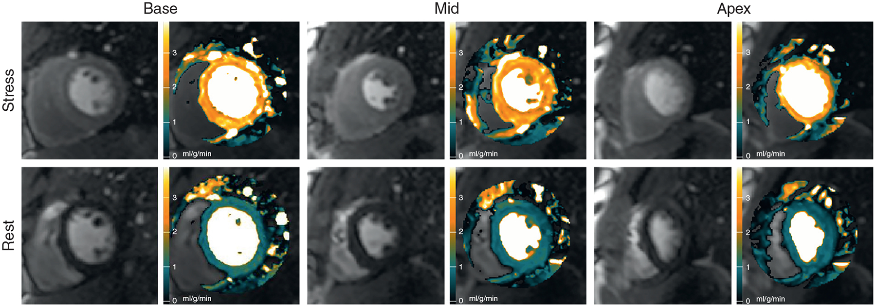
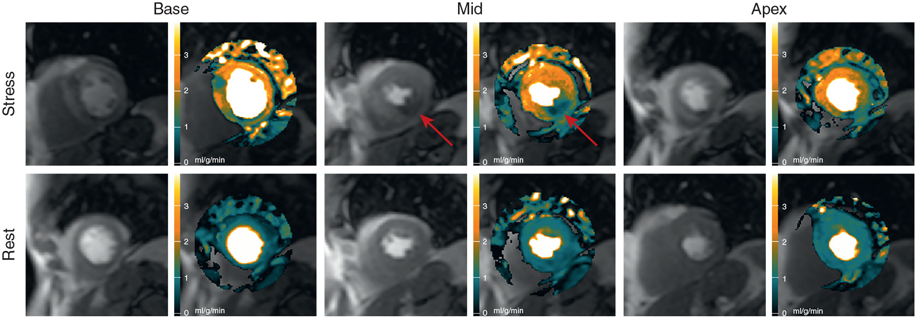
The correlation between automated and manual quantification was excellent (r = 0.96). Stress MBF and MPR in the ischemic zone were lower than those in the remote myocardium in patients with significant CAD (both p < 0.001). Stress MBF and MPR in the remote zone of the patients were lower than those in the normal volunteers (both p < 0.001). All quantitative metrics had good area under the curve (0.864 to 0.926), sensitivity (82.9% to 91.4%), and specificity (75.6% to 91.1%) on per-patient analysis. On a per-vessel analysis of the quantitative metrics, area under the curve (0.837 to 0.864), sensitivity (75.0% to 82.7%), and specificity (71.8% to 80.9%) were good.

CONCLUSIONS

Fully quantitative CMR MBF pixel maps can be generated automatically, and the results agree well with manual quantification. These methods can discriminate regional perfusion variations and have high diagnostic performance for detecting significant CAD. (Technical Development of Cardiovascular Magnetic Resonance Imaging; NCT00027170).

摘要

目的

作者开发了一种完全自动化的框架,用于从对比增强心脏磁共振(CMR)灌注成像中定量测量心肌血流(MBF),并评估其在患者中的诊断性能。

背景

以前已经通过微球 MBF 测量对完全定量的 CMR 灌注像素图进行了验证,并且显示出了在临床应用中的潜力,但是这些方法需要繁琐的手动过程,并且过于耗时。

方法

对 80 例已知或疑似冠状动脉疾病(CAD)患者和 17 例健康志愿者进行 CMR 灌注成像。定量冠状动脉造影(QCA)定义为≥70%狭窄的 CAD 为显著 CAD;QCA 定义为<70%狭窄或冠状动脉计算机断层血管造影术(CCTA)定义为<30%狭窄且所有血管的钙评分均为 0 的 CAD 为非显著 CAD。与健康志愿者的手动定量相比,自动生成的 MBF 图进行比较。使用绝对 MBF、心肌灌注储备(MPR)和 MBF 和 MPR 的相对测量值,对患者的自动 MBF 像素图的诊断性能进行分析。

结果

自动和手动定量之间的相关性很好(r=0.96)。有显著 CAD 的患者的缺血区的应激 MBF 和 MPR 均低于远隔心肌(均 p<0.001)。患者的远隔区的应激 MBF 和 MPR 均低于正常志愿者(均 p<0.001)。所有定量指标在患者的逐例分析中均具有良好的曲线下面积(0.864 至 0.926)、敏感性(82.9%至 91.4%)和特异性(75.6%至 91.1%)。在定量指标的逐支血管分析中,曲线下面积(0.837 至 0.864)、敏感性(75.0%至 82.7%)和特异性(71.8%至 80.9%)均较好。

结论

完全定量的 CMR MBF 像素图可以自动生成,并且结果与手动定量吻合良好。这些方法可以区分区域性灌注变化,对检测显著 CAD 具有较高的诊断性能。(心血管磁共振成像技术发展;NCT00027170)。

https://cdn.ncbi.nlm.nih.gov/pmc/blobs/694b/8760891/6d470d9a8707/nihms-968296-f0007.jpg
https://cdn.ncbi.nlm.nih.gov/pmc/blobs/694b/8760891/1041b171b8dc/nihms-968296-f0001.jpg
https://cdn.ncbi.nlm.nih.gov/pmc/blobs/694b/8760891/5c3b10590ff6/nihms-968296-f0002.jpg
https://cdn.ncbi.nlm.nih.gov/pmc/blobs/694b/8760891/bd1b3f949be8/nihms-968296-f0003.jpg
https://cdn.ncbi.nlm.nih.gov/pmc/blobs/694b/8760891/1f074495a90a/nihms-968296-f0004.jpg
https://cdn.ncbi.nlm.nih.gov/pmc/blobs/694b/8760891/49e261a45480/nihms-968296-f0005.jpg
https://cdn.ncbi.nlm.nih.gov/pmc/blobs/694b/8760891/051a97669c5f/nihms-968296-f0006.jpg
https://cdn.ncbi.nlm.nih.gov/pmc/blobs/694b/8760891/6d470d9a8707/nihms-968296-f0007.jpg

相似文献

1
Diagnostic Performance of Fully Automated Pixel-Wise Quantitative Myocardial Perfusion Imaging by Cardiovascular Magnetic Resonance.心血管磁共振全自动像素定量心肌灌注成像的诊断性能。
JACC Cardiovasc Imaging. 2018 May;11(5):697-707. doi: 10.1016/j.jcmg.2018.01.005. Epub 2018 Feb 14.
2
Fully automated pixel-wise quantitative CMR-myocardial perfusion with CMR-coronary angiography to detect hemodynamically significant coronary artery disease.应用 CMR 冠状动脉造影全自动像素级定量 CMR 心肌灌注成像检测有血流动力学意义的冠状动脉疾病。
Eur Radiol. 2023 Oct;33(10):7238-7249. doi: 10.1007/s00330-023-09689-8. Epub 2023 May 5.
3
Fully quantitative pixel-wise analysis of cardiovascular magnetic resonance perfusion improves discrimination of dark rim artifact from perfusion defects associated with epicardial coronary stenosis.对心血管磁共振灌注的完全定量像素分析可改善对与心外膜冠状动脉狭窄相关的暗带伪影与灌注缺损的区分。
J Cardiovasc Magn Reson. 2018 Mar 8;20(1):16. doi: 10.1186/s12968-018-0436-0.
4
Automated Pixel-Wise Quantitative Myocardial Perfusion Mapping by CMR to Detect Obstructive Coronary Artery Disease and Coronary Microvascular Dysfunction: Validation Against Invasive Coronary Physiology.CMR 自动像素定量心肌灌注成像检测冠状动脉疾病和冠状动脉微血管功能障碍:与有创性冠状动脉生理学的对照验证。
JACC Cardiovasc Imaging. 2019 Oct;12(10):1958-1969. doi: 10.1016/j.jcmg.2018.12.022. Epub 2019 Feb 13.
5
Quantitative three-dimensional cardiovascular magnetic resonance myocardial perfusion imaging in systole and diastole.定量三维心血管磁共振心动周期收缩期和舒张期心肌灌注成像。
J Cardiovasc Magn Reson. 2014 Feb 24;16(1):19. doi: 10.1186/1532-429X-16-19.
6
Diagnostic value of global myocardial perfusion reserve assessment based on coronary sinus flow measurements using cardiovascular magnetic resonance in addition to myocardial stress perfusion imaging.基于冠状动脉窦血流测量的心肌整体灌注储备评估在心肌负荷灌注成像之外的诊断价值。
Eur Heart J Cardiovasc Imaging. 2017 May 1;18(8):851-859. doi: 10.1093/ehjci/jew315.
7
A comparison of cardiovascular magnetic resonance and single photon emission computed tomography (SPECT) perfusion imaging in left main stem or equivalent coronary artery disease: a CE-MARC substudy.左主干或等效冠状动脉疾病中心血管磁共振与单光子发射计算机断层扫描(SPECT)灌注成像的比较:CE-MARC 子研究。
J Cardiovasc Magn Reson. 2017 Nov 6;19(1):84. doi: 10.1186/s12968-017-0398-7.
8
Fully automated, inline quantification of myocardial blood flow with cardiovascular magnetic resonance: repeatability of measurements in healthy subjects.使用心血管磁共振进行全自动、在线心肌血流定量:健康受试者测量的可重复性。
J Cardiovasc Magn Reson. 2018 Jul 9;20(1):48. doi: 10.1186/s12968-018-0462-y.
9
Comparison of machine learning-based CT fractional flow reserve with cardiac MR perfusion mapping for ischemia diagnosis in stable coronary artery disease.基于机器学习的 CT 分数血流量储备与心脏磁共振灌注成像在稳定型冠状动脉疾病缺血诊断中的比较。
Eur Radiol. 2024 Sep;34(9):5654-5665. doi: 10.1007/s00330-024-10650-6. Epub 2024 Feb 26.
10
A quantitative pixel-wise measurement of myocardial blood flow by contrast-enhanced first-pass CMR perfusion imaging: microsphere validation in dogs and feasibility study in humans.对比增强首过 CMR 灌注成像定量像素级心肌血流测量:微球在犬中的验证和在人体中的可行性研究。
JACC Cardiovasc Imaging. 2012 Feb;5(2):154-66. doi: 10.1016/j.jcmg.2011.07.013.

引用本文的文献

1
Next-Generation Cardiac Magnetic Resonance Imaging Techniques for Characterization of Myocardial Disease.用于心肌疾病特征描述的下一代心脏磁共振成像技术
Curr Treat Options Cardiovasc Med. 2024 Aug;26(8):243-254. doi: 10.1007/s11936-024-01044-4. Epub 2024 Aug 9.
2
Advanced Cardiac Magnetic Resonance Imaging for Assessment of Obstructive Coronary Artery Disease - ADVOCATE-CMR Study Rationale and Design.用于评估阻塞性冠状动脉疾病的高级心脏磁共振成像——ADVOCATE-CMR研究原理与设计
J Cardiovasc Magn Reson. 2025 Apr 25:101900. doi: 10.1016/j.jocmr.2025.101900.
3
Prevalence and prognostic significance of reduced myocardial perfusion reserve in diabetic heart failure with preserved ejection fraction using quantitative perfusion cardiac magnetic resonance.

本文引用的文献

1
Diagnostic performance of semi-quantitative and quantitative stress CMR perfusion analysis: a meta-analysis.半定量和定量压力 CMR 灌注分析的诊断性能:荟萃分析。
J Cardiovasc Magn Reson. 2017 Nov 27;19(1):92. doi: 10.1186/s12968-017-0393-z.
2
Myocardial perfusion cardiovascular magnetic resonance: optimized dual sequence and reconstruction for quantification.心肌灌注心血管磁共振成像:用于定量分析的优化双序列及重建方法
J Cardiovasc Magn Reson. 2017 Apr 7;19(1):43. doi: 10.1186/s12968-017-0355-5.
3
Robust universal nonrigid motion correction framework for first-pass cardiac MR perfusion imaging.
使用定量灌注心脏磁共振成像评估射血分数保留的糖尿病心力衰竭患者心肌灌注储备降低的患病率及预后意义
Eur Radiol. 2025 Mar 5. doi: 10.1007/s00330-025-11474-8.
4
Quantitative perfusion by cardiac magnetic resonance imaging reveals compromised myocardial perfusion in patients with angina with non-obstructive coronary artery disease.心脏磁共振成像定量灌注显示,非阻塞性冠状动脉疾病所致心绞痛患者存在心肌灌注受损。
Clin Res Cardiol. 2025 Feb 18. doi: 10.1007/s00392-025-02606-7.
5
Stress T1 mapping and quantitative perfusion cardiovascular magnetic resonance in patients with suspected obstructive coronary artery disease.疑似阻塞性冠状动脉疾病患者的应力T1映射和定量灌注心血管磁共振成像
Eur Heart J Cardiovasc Imaging. 2025 May 30;26(6):980-990. doi: 10.1093/ehjci/jeaf059.
6
Artificial Intelligence in the Heart of Medicine: A Systematic Approach to Transforming Arrhythmia Care with Intelligent Systems.医学核心领域的人工智能:利用智能系统转变心律失常护理的系统方法。
Curr Cardiol Rev. 2025;21(4):e1573403X334095. doi: 10.2174/011573403X334095241205041550.
7
Heart-brain microvascular MRI study: protocol for a multicentre, observational, cohort study in the UK assessing associations between small vessel disease of the heart and brain.心脏-脑微血管磁共振成像研究:英国一项多中心、观察性队列研究的方案,评估心脏和脑小血管疾病之间的关联。
BMJ Open. 2024 Dec 20;14(12):e088372. doi: 10.1136/bmjopen-2024-088372.
8
Assessing microvascular dysfunction and predicting long-term prognosis in patients with cardiac amyloidosis by cardiovascular magnetic resonance quantitative stress perfusion.通过心血管磁共振定量应力灌注评估心脏淀粉样变性患者的微血管功能障碍并预测其长期预后。
J Cardiovasc Magn Reson. 2024 Dec 14;27(1):101134. doi: 10.1016/j.jocmr.2024.101134.
9
Diagnostic performance of quantitative perfusion cardiac magnetic resonance imaging in patients with prior coronary artery disease.定量灌注心脏磁共振成像在既往有冠状动脉疾病患者中的诊断性能
Eur Heart J Cardiovasc Imaging. 2025 Jan 31;26(2):207-217. doi: 10.1093/ehjci/jeae262.
10
Coronary microvascular dysfunction in autoimmune rheumatic diseases: beyond coronary flow velocity reserve.自身免疫性风湿性疾病中的冠状动脉微血管功能障碍:超越冠状动脉血流储备
Front Cardiovasc Med. 2024 Aug 21;11:1372703. doi: 10.3389/fcvm.2024.1372703. eCollection 2024.
用于首过心脏磁共振灌注成像的稳健通用非刚性运动校正框架。
J Magn Reson Imaging. 2017 Oct;46(4):1060-1072. doi: 10.1002/jmri.25659. Epub 2017 Feb 15.
4
Evaluation of an automated method for arterial input function detection for first-pass myocardial perfusion cardiovascular magnetic resonance.用于首次通过心肌灌注心血管磁共振的动脉输入函数检测自动化方法的评估。
J Cardiovasc Magn Reson. 2016 Apr 8;18:17. doi: 10.1186/s12968-016-0239-0.
5
Splenic Switch-off: A Tool to Assess Stress Adequacy in Adenosine Perfusion Cardiac MR Imaging.脾脏失活:评估腺苷灌注心脏磁共振成像中应激适度的工具。
Radiology. 2015 Sep;276(3):732-40. doi: 10.1148/radiol.2015142059. Epub 2015 Apr 29.
6
Multicenter evaluation of dynamic three-dimensional magnetic resonance myocardial perfusion imaging for the detection of coronary artery disease defined by fractional flow reserve.多中心评估动态三维磁共振心肌灌注成像在检测以血流储备分数定义的冠状动脉疾病中的应用。
Circ Cardiovasc Imaging. 2015 May;8(5). doi: 10.1161/CIRCIMAGING.114.003061.
7
FLASH proton density imaging for improved surface coil intensity correction in quantitative and semi-quantitative SSFP perfusion cardiovascular magnetic resonance.用于定量和半定量稳态自由进动灌注心血管磁共振成像中改善表面线圈强度校正的快速质子密度成像。
J Cardiovasc Magn Reson. 2015 Feb 17;17(1):16. doi: 10.1186/s12968-015-0120-6.
8
Comparison of the Diagnostic Performance of Four Quantitative Myocardial Perfusion Estimation Methods Used in Cardiac MR Imaging: CE-MARC Substudy.心脏磁共振成像中四种定量心肌灌注估计方法的诊断性能比较:CE-MARC子研究
Radiology. 2015 May;275(2):393-402. doi: 10.1148/radiol.14140433. Epub 2014 Dec 18.
9
Coronary microvascular ischemia in hypertrophic cardiomyopathy - a pixel-wise quantitative cardiovascular magnetic resonance perfusion study.肥厚型心肌病中的冠状动脉微血管缺血——一项逐像素定量心血管磁共振灌注研究
J Cardiovasc Magn Reson. 2014 Aug 12;16(1):49. doi: 10.1186/s12968-014-0049-1.
10
Voxel-wise quantification of myocardial blood flow with cardiovascular magnetic resonance: effect of variations in methodology and validation with positron emission tomography.利用心血管磁共振进行心肌血流的体素定量分析:方法学差异的影响及正电子发射断层扫描验证
J Cardiovasc Magn Reson. 2014 Jan 24;16(1):11. doi: 10.1186/1532-429X-16-11.