Dumontel B, Canta M, Engelke H, Chiodoni A, Racca L, Ancona A, Limongi T, Canavese G, Cauda V
Department of Applied Science and Technology , Politecnico di Torino , Corso Duca degli Abruzzi 24 , 10129 Turin , Italy . Email:
Department of Chemistry , Ludwig-Maximilians-University of Munich , Butenandtstrasse 11E , 81377 Munich , Germany.
J Mater Chem B. 2017 Nov 28;5(44):8799-8813. doi: 10.1039/c7tb02229h. Epub 2017 Nov 1.
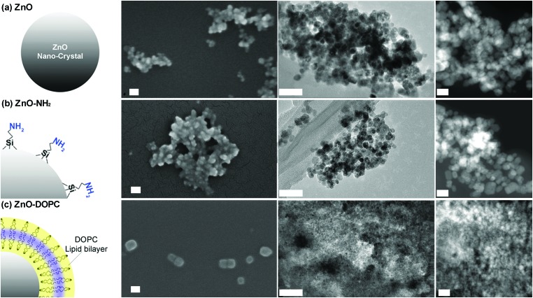
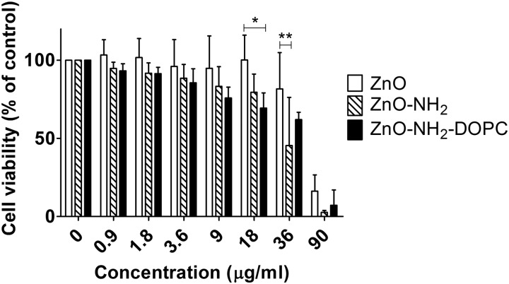
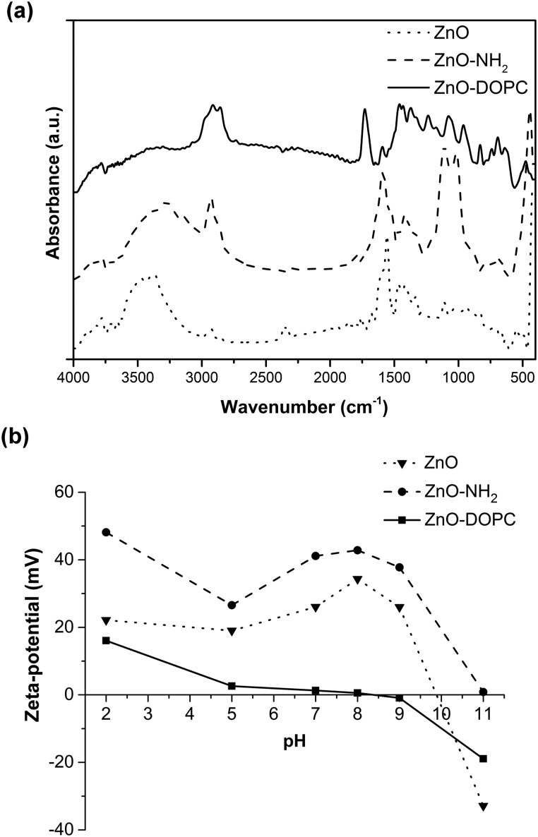
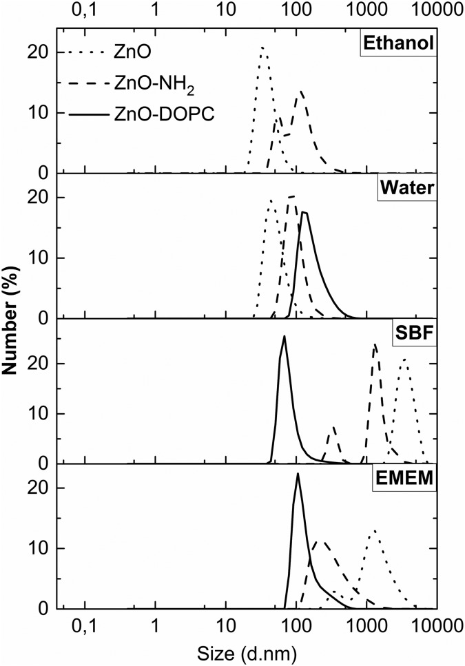
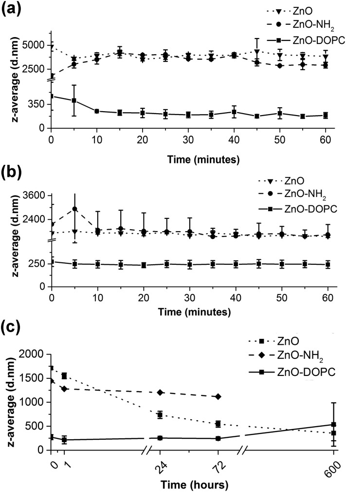
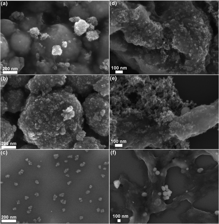
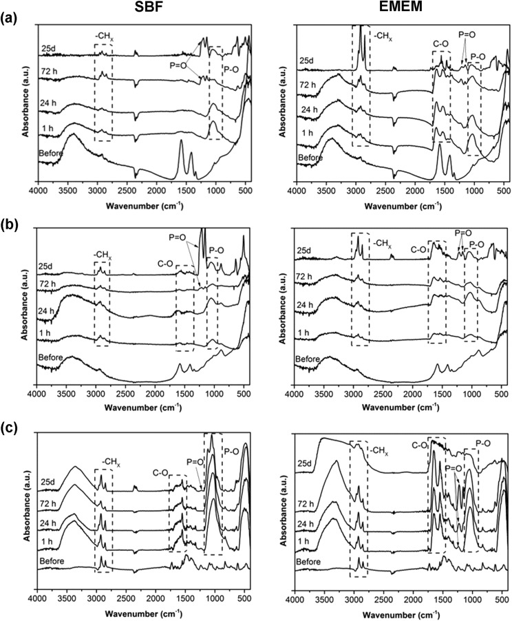
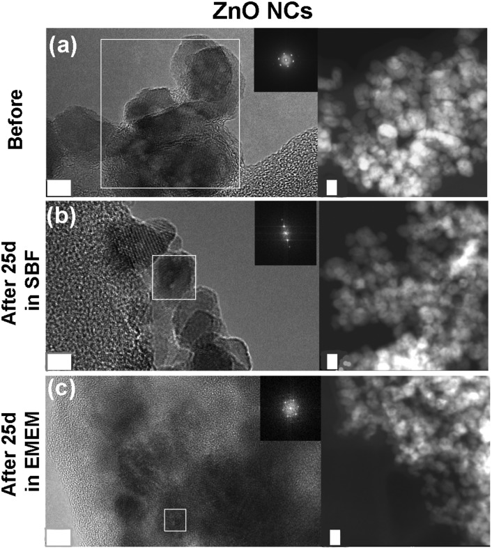
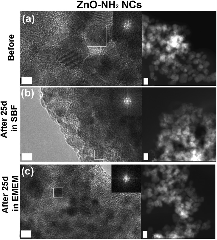
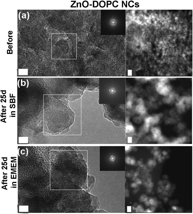
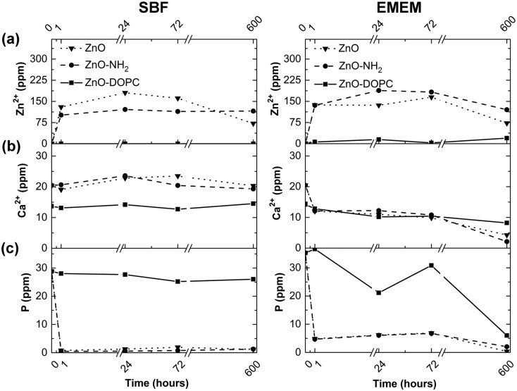
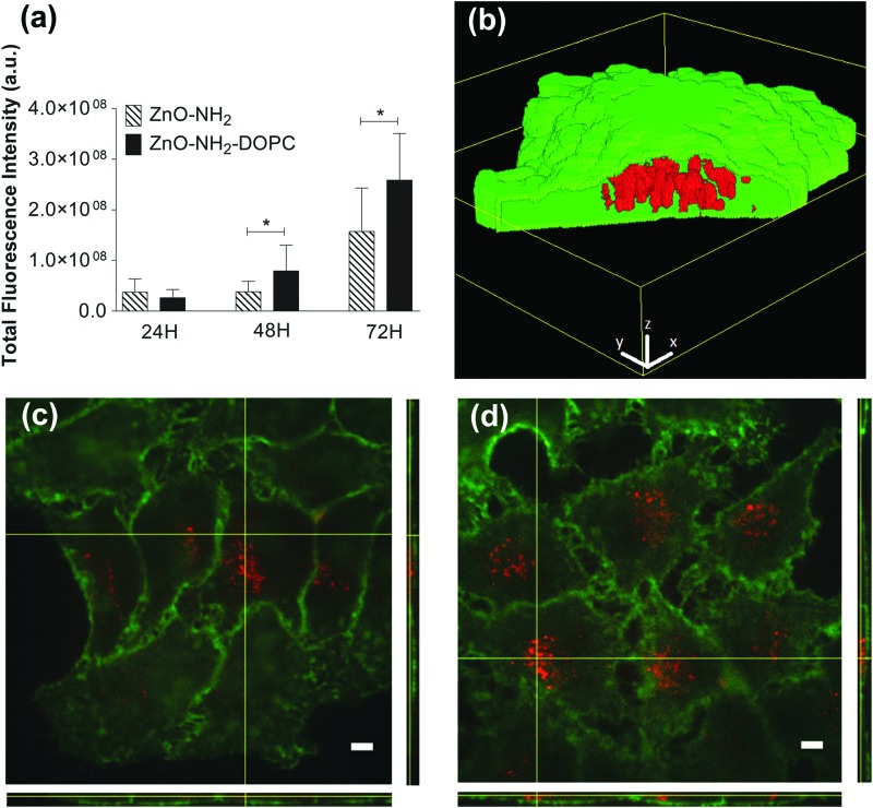
The widespread use of ZnO nanomaterials for biomedical applications, including therapeutic drug delivery or stimuli-responsive activation, as well as imaging, imposes a careful control over the colloidal stability and long-term behaviour of ZnO in biological media. Moreover, the effect of ZnO nanostructures on living cells, in particular cancer cells, is still under debate. This paper discusses the role of surface chemistry and charge of zinc oxide nanocrystals, of around 15 nm in size, which influence their behaviour in biological fluids and effect on cancer cells. In particular, we address this problem by modifying the surface of pristine ZnO nanocrystals (NCs), rich of hydroxyl groups, with positively charged amino-propyl chains or, more innovatively, by self-assembling a double-lipidic membrane, shielding the ZnO NCs. Our findings show that the prolonged immersion in simulated human plasma and in the cell culture medium leads to highly colloidally dispersed ZnO NCs only when coated by the lipidic bilayer. In contrast, the pristine and amine-functionalized NCs form huge aggregates after already one hour of immersion. Partial dissolution of these two samples into potentially cytotoxic Zn cations takes place, together with the precipitation of phosphate and carbonate salts on the NCs' surface. When exposed to living HeLa cancer cells, higher amounts of lipid-shielded ZnO NCs are internalized with respect to the other samples, thus showing a reduced cytotoxicity, based on the same amount of internalized NCs. These results pave the way for the development of novel theranostic platforms based on ZnO NCs. The new formulation of ZnO shielded with a lipid-bilayer will prevent strong aggregation and premature degradation into toxic by-products, and promote a highly efficient cell uptake for further therapeutic or diagnostic functions.
氧化锌纳米材料在生物医学领域有着广泛应用,包括治疗药物递送、刺激响应激活以及成像等,这就要求对氧化锌在生物介质中的胶体稳定性和长期行为进行严格控制。此外,氧化锌纳米结构对活细胞,尤其是癌细胞的影响仍存在争议。本文讨论了尺寸约为15纳米的氧化锌纳米晶体的表面化学性质和电荷的作用,它们会影响纳米晶体在生物流体中的行为以及对癌细胞的影响。具体而言,我们通过用带正电荷的氨基丙基链修饰富含羟基的原始氧化锌纳米晶体(NCs)表面,或者更具创新性地通过自组装双层脂质膜来屏蔽氧化锌纳米晶体,从而解决这一问题。我们的研究结果表明,只有当用脂质双层包覆时,长时间浸泡在模拟人体血浆和细胞培养基中才会使氧化锌纳米晶体高度胶体分散。相比之下,原始的和胺功能化的纳米晶体在浸泡仅一小时后就会形成巨大的聚集体。这两个样品会部分溶解为潜在的细胞毒性锌阳离子,同时磷酸盐和碳酸盐盐会在纳米晶体表面沉淀。当暴露于活的HeLa癌细胞时,相对于其他样品,更多的脂质屏蔽氧化锌纳米晶体会被内化,因此基于相同数量的内化纳米晶体,其细胞毒性降低。这些结果为基于氧化锌纳米晶体的新型治疗诊断平台的开发铺平了道路。用脂质双层屏蔽的氧化锌新配方将防止强烈聚集和过早降解为有毒副产物,并促进高效的细胞摄取以实现进一步的治疗或诊断功能。