Department of Chemistry , University of California , Berkeley , California 94720 , United States.
DOE Joint Genome Institute , 2800 Mitchell Drive , Walnut Creek , California 94598 , United States.
ACS Chem Biol. 2018 Jul 20;13(7):1872-1879. doi: 10.1021/acschembio.7b01019. Epub 2018 Mar 5.
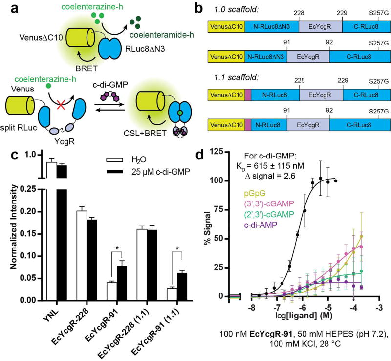
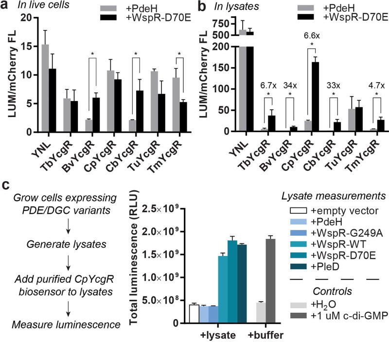
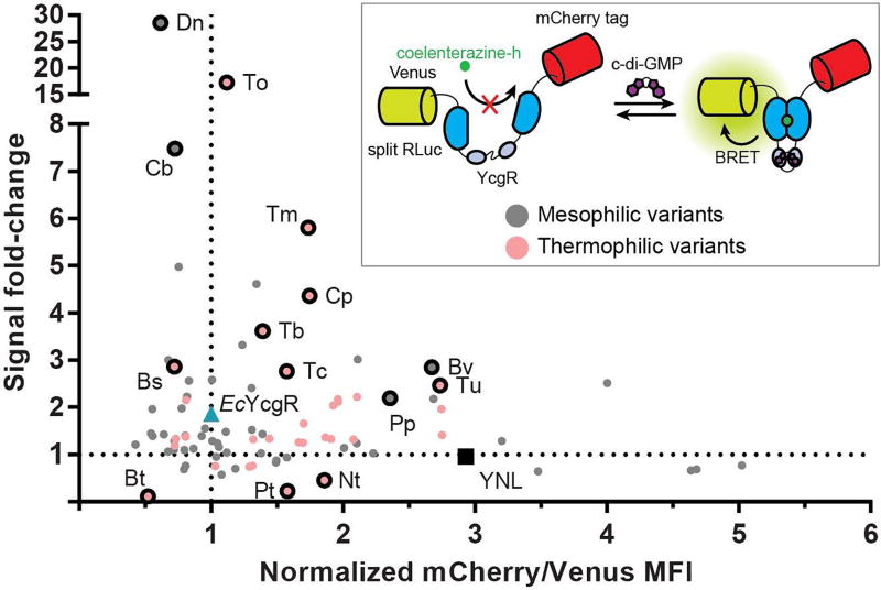
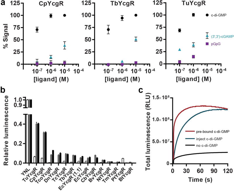
Bacteria colonize highly diverse and complex environments, from gastrointestinal tracts to soil and plant surfaces. This colonization process is controlled in part by the intracellular signal cyclic di-GMP, which regulates bacterial motility and biofilm formation. To interrogate cyclic di-GMP signaling networks, a variety of fluorescent biosensors for live cell imaging of cyclic di-GMP have been developed. However, the need for external illumination precludes the use of these tools for imaging bacteria in their natural environments, including in deep tissues of whole organisms and in samples that are highly autofluorescent or photosensitive. The need for genetic encoding also complicates the analysis of clinical isolates and environmental samples. Toward expanding the study of bacterial signaling to these systems, we have developed the first chemiluminescent biosensors for cyclic di-GMP. The biosensor design combines the complementation of split luciferase (CSL) and bioluminescence resonance energy transfer (BRET) approaches. Furthermore, we developed a lysate-based assay for biosensor activity that enabled reliable high-throughput screening of a phylogenetic library of 92 biosensor variants. The screen identified biosensors with very large signal changes (∼40- and 90-fold) as well as biosensors with high affinities for cyclic di-GMP ( K < 50 nM). These chemiluminescent biosensors then were applied to measure cyclic di-GMP levels in E. coli. The cellular experiments revealed an unexpected challenge for chemiluminescent imaging in Gram negative bacteria but showed promising application in lysates. Taken together, this work establishes the first chemiluminescent biosensors for studying cyclic di-GMP signaling and provides a foundation for using these biosensors in more complex systems.
细菌栖息于多样化且复杂的环境中,从胃肠道到土壤和植物表面。这一栖息过程受到胞内信号分子环二鸟苷酸(cyclic di-GMP)的部分调控,它调节着细菌的运动性和生物被膜的形成。为了研究环二鸟苷酸信号网络,人们开发了多种用于环二鸟苷酸活细胞成像的荧光生物传感器。然而,这些工具需要外部光照,因此无法用于在自然环境中对细菌进行成像,包括在整个生物体的深部组织中以及在高度自发荧光或对光敏感的样本中。对遗传编码的需求也使临床分离株和环境样本的分析变得复杂。为了将细菌信号的研究扩展到这些系统,我们开发了首个用于环二鸟苷酸的化学发光生物传感器。该生物传感器设计结合了分裂荧光酶(split luciferase,CSL)和生物发光共振能量转移(bioluminescence resonance energy transfer,BRET)方法。此外,我们开发了一种基于裂解物的生物传感器活性测定方法,可用于对 92 种生物传感器变体的系统发育文库进行可靠的高通量筛选。该筛选确定了具有非常大信号变化(约 40 倍和 90 倍)的生物传感器以及对环二鸟苷酸具有高亲和力( K < 50 nM)的生物传感器。然后,这些化学发光生物传感器被用于测量大肠杆菌中的环二鸟苷酸水平。细胞实验揭示了革兰氏阴性菌中化学发光成像的一个意外挑战,但在裂解物中显示出了有前景的应用。总之,这项工作建立了首个用于研究环二鸟苷酸信号的化学发光生物传感器,并为在更复杂的系统中使用这些生物传感器提供了基础。