Robson Annie-Louise, Dastoor Paul C, Flynn Jamie, Palmer William, Martin Antony, Smith Doug W, Woldu Ameha, Hua Susan
School of Biomedical Sciences and Pharmacy, University of Newcastle, Callaghan, NSW, Australia.
Centre for Organic Electronics, University of Newcastle, Callaghan, NSW, Australia.
Front Pharmacol. 2018 Feb 6;9:80. doi: 10.3389/fphar.2018.00080. eCollection 2018.
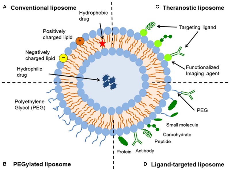
There are currently a number of imaging techniques available for evaluating the morphology of liposomes and other nanoparticles, with each having its own advantages and disadvantages that should be considered when interpreting data. Controlling and validating the morphology of nanoparticles is of key importance for the effective clinical translation of liposomal formulations. There are a number of physical characteristics of liposomes that determine their behavior, including size, surface characteristics, lamellarity, and homogeneity. Despite the great importance of the morphology of nanoparticles, it is generally not well-characterized and is difficult to control. Appropriate imaging techniques provide important details regarding the morphological characteristics of nanoparticles, and should be used in conjunction with other methods to assess physicochemical parameters. In this review, we will discuss the advantages and limitations of available imaging techniques used to evaluate liposomal formulations.
目前有多种成像技术可用于评估脂质体和其他纳米颗粒的形态,每种技术都有其自身的优缺点,在解释数据时应予以考虑。控制和验证纳米颗粒的形态对于脂质体制剂的有效临床转化至关重要。脂质体有许多决定其行为的物理特性,包括大小、表面特性、层数和均匀性。尽管纳米颗粒的形态非常重要,但它通常没有得到很好的表征且难以控制。合适的成像技术可提供有关纳米颗粒形态特征的重要细节,并且应与其他方法结合使用以评估物理化学参数。在本综述中,我们将讨论用于评估脂质体制剂的现有成像技术的优缺点。