• 文献检索
  • 文档翻译
  • 深度研究
  • 学术资讯
  • Suppr Zotero 插件Zotero 插件
  • 邀请有礼
  • 套餐&价格
  • 历史记录
应用&插件
Suppr Zotero 插件Zotero 插件浏览器插件Mac 客户端Windows 客户端微信小程序
定价
高级版会员购买积分包购买API积分包
服务
文献检索文档翻译深度研究API 文档MCP 服务
关于我们
关于 Suppr公司介绍联系我们用户协议隐私条款
关注我们

Suppr 超能文献

核心技术专利:CN118964589B侵权必究
粤ICP备2023148730 号-1Suppr @ 2026

文献检索

告别复杂PubMed语法,用中文像聊天一样搜索,搜遍4000万医学文献。AI智能推荐,让科研检索更轻松。

立即免费搜索

文件翻译

保留排版,准确专业,支持PDF/Word/PPT等文件格式,支持 12+语言互译。

免费翻译文档

深度研究

AI帮你快速写综述,25分钟生成高质量综述,智能提取关键信息,辅助科研写作。

立即免费体验

特发性阴囊钙质沉着症——病例报告

Idiopathic scrotal calcinosis - A case report.

作者信息

Tareen Ali, Ibrahim Rami Mossad

机构信息

Dept. of Dermatology, Odense University Hospital, Odense C, Denmark.

Dept. of Plasticsurgery, Herlev Hospital, Copenhagen, Denmark.

出版信息

Int J Surg Case Rep. 2018;44:51-53. doi: 10.1016/j.ijscr.2018.02.010. Epub 2018 Feb 15.

DOI:10.1016/j.ijscr.2018.02.010
PMID:29477103
原文链接:https://pmc.ncbi.nlm.nih.gov/articles/PMC5928033/
Abstract

INTRODUCTION

Idiopathic scrotal calcinosis is a rare benign condition which presents with asymptomatic multiple nodules on the scrotal skin.

PRESENTATION OF CASE

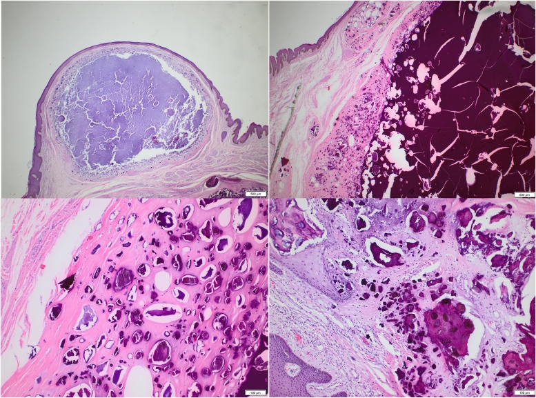
Our patient, a 64-year-old Indian male with Fitzpatrick skin type 4, presented with multiple nodules, which were completely surgically excised with no complications. Histological examination reveals extensive intradermal deposition of calcium surrounded by histiocytes and without cystic structure.

DISCUSSION

Numerous theories about the pathogenesis have been proposed and the evidence presented suggests this is a continuum.

CONCLUSION

The nature of idiopathic scrotal calcinosis is still unknown and it is up to debate whether the term "idiopathic" is appropriate for the condition.

摘要

引言

特发性阴囊钙化是一种罕见的良性疾病,表现为阴囊皮肤出现无症状的多个结节。

病例介绍

我们的患者是一名64岁的印度男性, Fitzpatrick皮肤类型为4型,出现多个结节,通过手术将其完全切除,无并发症。组织学检查显示真皮内有广泛的钙沉积,周围有组织细胞,无囊性结构。

讨论

关于发病机制已经提出了许多理论,现有证据表明这是一个连续过程。

结论

特发性阴囊钙化的本质仍然未知,“特发性”这个术语是否适用于这种情况仍有待争论。

https://cdn.ncbi.nlm.nih.gov/pmc/blobs/9e23/5928033/09eddbf50aa8/gr2.jpg
https://cdn.ncbi.nlm.nih.gov/pmc/blobs/9e23/5928033/0eda1a2f5759/gr1.jpg
https://cdn.ncbi.nlm.nih.gov/pmc/blobs/9e23/5928033/09eddbf50aa8/gr2.jpg
https://cdn.ncbi.nlm.nih.gov/pmc/blobs/9e23/5928033/0eda1a2f5759/gr1.jpg
https://cdn.ncbi.nlm.nih.gov/pmc/blobs/9e23/5928033/09eddbf50aa8/gr2.jpg

相似文献

1
Idiopathic scrotal calcinosis - A case report.特发性阴囊钙质沉着症——病例报告
Int J Surg Case Rep. 2018;44:51-53. doi: 10.1016/j.ijscr.2018.02.010. Epub 2018 Feb 15.
2
Scrotal calcinosis effect - Case report and review of literature.阴囊钙质沉着症的影响——病例报告及文献综述
Urol Case Rep. 2020 Jul 18;33:101347. doi: 10.1016/j.eucr.2020.101347. eCollection 2020 Nov.
3
ULTRASOUND IMAGING AND PATHOLOGIC FEATURES OF IDIOPATHIC SCROTAL CALCINOSIS IN A YOUNG ADULT NIGERIAN AND REVIEW OF THE LITERATURE.一名年轻尼日利亚成年人特发性阴囊钙化的超声成像与病理特征及文献综述
Ann Ib Postgrad Med. 2024 Apr 30;22(1):112-115.
4
Idiopathic Scrotal Calcinosis.特发性阴囊钙化症
Indian J Surg. 2016 Aug;78(4):329-30. doi: 10.1007/s12262-016-1463-4. Epub 2016 Mar 19.
5
Idiopathic scrotal calcinosis: the incorrect terminology of scrotal calcinosis.特发性阴囊钙化症:阴囊钙化症的错误术语。
G Ital Dermatol Venereol. 2015 Oct;150(5):495-9.
6
Idiopathic scrotal calcinosis: Is the cause still unknown? A case report.特发性阴囊钙化症:病因仍不明?一例报告。
Int J Surg Case Rep. 2024 Apr;117:109503. doi: 10.1016/j.ijscr.2024.109503. Epub 2024 Mar 11.
7
Scrotal calcinosis: idiopathic or dystrophic?阴囊钙质沉着症:特发性还是营养不良性?
Dermatol Online J. 2010 Feb 15;16(2):5.
8
Idiopathic scrotal calcinosis: report of 2 cases, and review of pathogenesis and factors that determine patients' acceptance of surgical treatment.特发性阴囊钙化症:2例报告及发病机制与决定患者接受手术治疗因素的文献复习
Clin Cosmet Investig Dermatol. 2018 Jul 11;11:333-337. doi: 10.2147/CCID.S142101. eCollection 2018.
9
Scrotal calcinosis due to resorption of cyst walls: a case report.囊肿壁吸收导致的阴囊钙化症:一例报告。
J Med Case Rep. 2008 Dec 8;2:375. doi: 10.1186/1752-1947-2-375.
10
Idiopathic Scrotal Calcinosis: A Case Report of a Rare Entity.特发性阴囊钙化症:一例罕见病例报告
Case Rep Urol. 2019 Dec 16;2019:6501964. doi: 10.1155/2019/6501964. eCollection 2019.

引用本文的文献

1
Young Man with Multiple Scrotal Growths.患有多个阴囊肿物的青年男性。
Oman Med J. 2022 Jul 31;37(4):e398. doi: 10.5001/omj.2022.04. eCollection 2022 Jul.
2
Idiopathic scrotal calcinosis: A new case report.特发性阴囊钙化症:一例新病例报告。
Urol Case Rep. 2020 Apr 25;32:101225. doi: 10.1016/j.eucr.2020.101225. eCollection 2020 Sep.

本文引用的文献

1
The SCARE Statement: Consensus-based surgical case report guidelines.SCARE 声明:基于共识的外科手术病例报告指南。
Int J Surg. 2016 Oct;34:180-186. doi: 10.1016/j.ijsu.2016.08.014. Epub 2016 Sep 7.
2
Idiopathic scrotal calcinosis: A rare entity and a review of the literature.特发性阴囊钙质沉着症:一种罕见病症及文献综述
Can Urol Assoc J. 2013 May-Jun;7(5-6):E439-41. doi: 10.5489/cuaj.1387.
3
Idiopathic scrotal calcinosis: report of two cases and review of literature.特发性阴囊钙化症:两例报告并文献复习
Int J Dermatol. 2012 Feb;51(2):199-203. doi: 10.1111/j.1365-4632.2011.04922.x. Epub 2011 Jul 5.
4
Calcinosis cutis: part I. Diagnostic pathway.皮肤钙化病:第一部分。诊断途径。
J Am Acad Dermatol. 2011 Jul;65(1):1-12; quiz 13-4. doi: 10.1016/j.jaad.2010.08.038.
5
Scrotal calcinosis: idiopathic or dystrophic?阴囊钙质沉着症:特发性还是营养不良性?
Dermatol Online J. 2010 Feb 15;16(2):5.
6
Risk factors associated with calcinosis of juvenile dermatomyositis.与青少年皮肌炎钙质沉着相关的危险因素。
J Pediatr (Rio J). 2008 Jan-Feb;84(1):68-74. doi: 10.2223/JPED.1746. Epub 2008 Jan 9.
7
Scrotal calcinosis results from calcification of cysts derived from hair follicles: a series of 20 cases evaluating the spectrum of changes resulting in scrotal calcinosis.阴囊钙化症是由毛囊衍生囊肿的钙化引起的:一项评估导致阴囊钙化症变化范围的20例系列研究。
Am J Dermatopathol. 2007 Apr;29(2):172-5. doi: 10.1097/01.dad.0000246465.25986.68.
8
Scrotal calcinosis: is the cause still unknown?阴囊钙质沉着症:病因仍不明?
J Am Acad Dermatol. 2004 Aug;51(2 Suppl):S97-S101. doi: 10.1016/j.jaad.2004.01.039.
9
An extremely severe case of cutaneous calcinosis with juvenile dermatomyositis, and successful treatment with diltiazem.一例伴有青少年皮肌炎的极其严重的皮肤钙化症,以及地尔硫卓成功治疗的病例。
Br J Dermatol. 2001 Apr;144(4):894-7. doi: 10.1046/j.1365-2133.2001.04153.x.
10
Head and neck pilomatricoma in the pediatric age group: a retrospective study and literature review.儿童年龄组头颈部毛发上皮瘤:一项回顾性研究及文献综述
Int J Pediatr Otorhinolaryngol. 2001 Feb;57(2):123-8. doi: 10.1016/s0165-5876(00)00449-3.