Department of Ultrasound, Renji Hospital, School of Medicine, Shanghai Jiao Tong University, 160 Pu Jian Rd, Shanghai, 200127, China.
Department of Biomedical Engineering, Case Western Reserve University Schools of Medicine, 10900 Euclid Ave., Cleveland, OH, 44106, USA.
Sci Rep. 2018 Mar 1;8(1):3887. doi: 10.1038/s41598-018-21502-x.
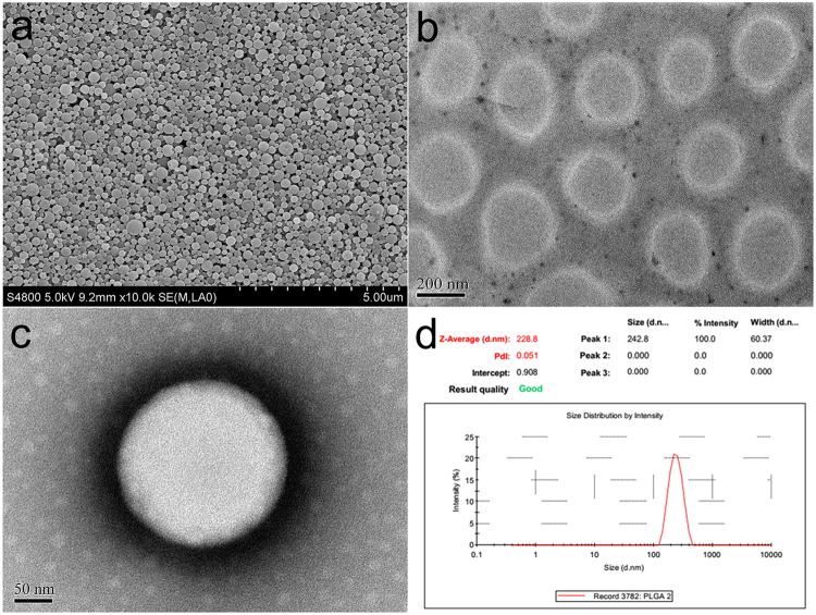
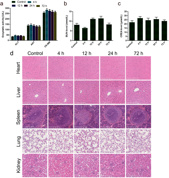
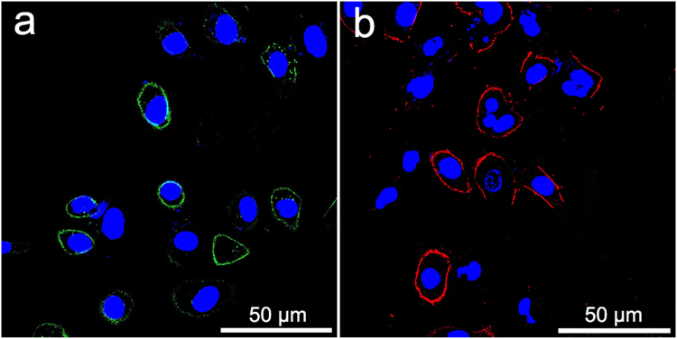
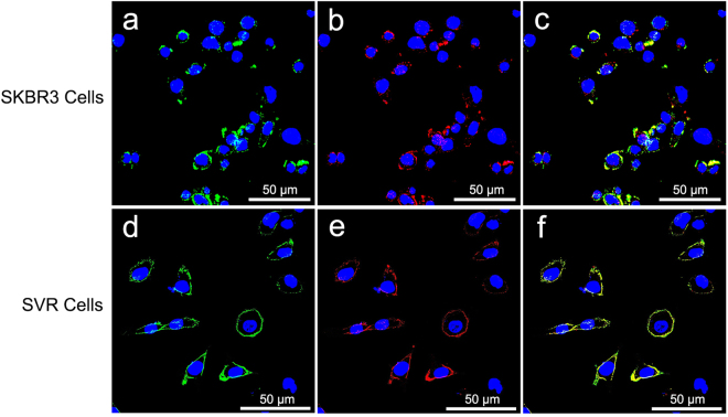
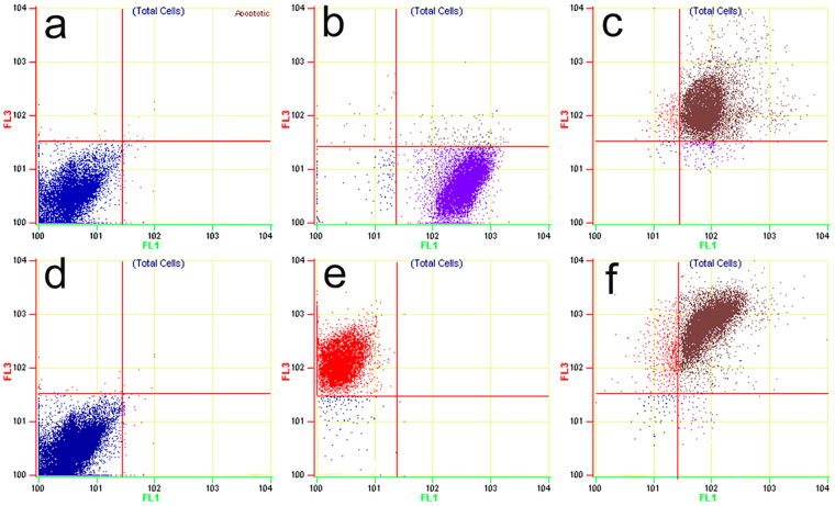
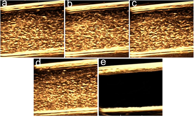
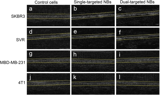
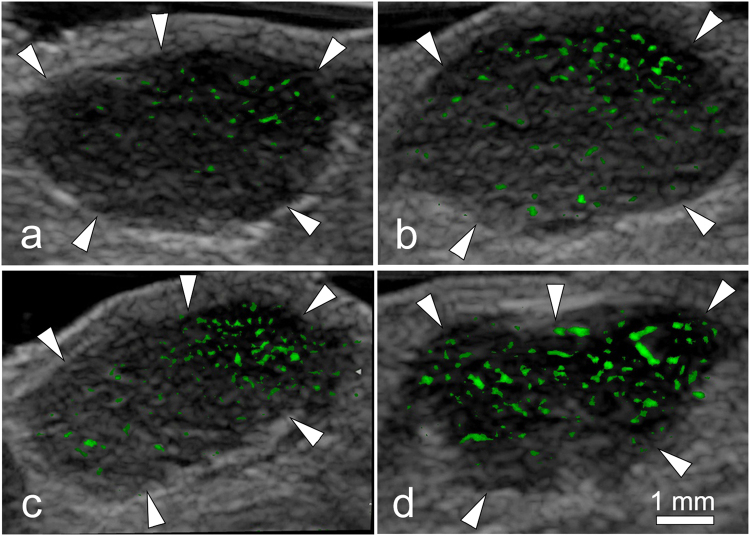
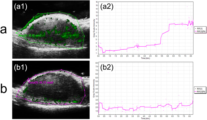
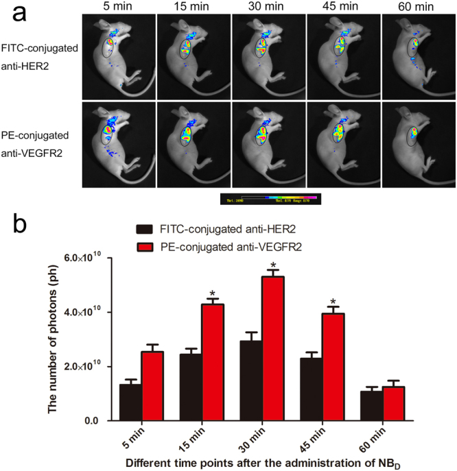
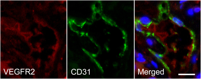
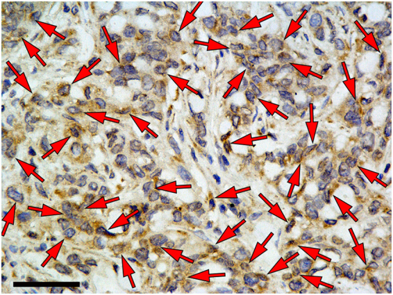
Molecularly-targeted contrast enhanced ultrasound (US) imaging is a promising imaging strategy with large potential for improving diagnostic accuracy of conventional US imaging in breast cancer detection. Therefore, we constructed a novel dual-targeted nanosized US contrast agent (UCA) directed at both vascular endothelial growth factor receptor 2 (VEGFR2) and human epidermal growth factor receptor 2 (HER2) based on perfluoropropane (CF)-filled poly(lactic-co-glycolic acid) (PLGA) (NBs) for breast cancer detection. In vitro, single- or dual-targeted PLGA NBs showed high target specificities and better effects of target enhancement in VEGFR2 or HER2-positive cells. In vivo, US imaging signal in the murine breast cancer model was significantly higher (P < 0.01) for dual-targeted NBs than single-targeted and non-targeted NBs. Small animal fluorescence imaging further confirmed the special affinity of the dual-targeted nanosized contrast agent to both VEGFR2 and HER2. Immunofluorescence and immunohistochemistry staining confirmed the expressions of VEGFR2 and HER2 on tumor neovasculature and tumor cells of breast cancer. In conclusions, the feasibility of using dual-targeted PLGA NBs to enhance ultrasonic images is demonstrated in vitro and in vivo. This may be a promising approach to target biomarkers of breast cancer for two site-specific US molecular imaging.
分子靶向对比增强超声(US)成像技术是一种很有前途的成像策略,具有很大的潜力,可以提高常规 US 成像在乳腺癌检测中的诊断准确性。因此,我们基于全氟丙烷(CF)填充的聚乳酸-羟基乙酸共聚物(PLGA)(NBs)构建了一种新型的双靶向纳米级超声对比剂(UCA),用于检测乳腺癌,该对比剂针对血管内皮生长因子受体 2(VEGFR2)和人表皮生长因子受体 2(HER2)。体外,单靶向或双靶向 PLGA NBs 对 VEGFR2 或 HER2 阳性细胞具有较高的靶向特异性和更好的靶向增强效果。在体内,与单靶向和非靶向 NBs 相比,双靶向 NBs 在小鼠乳腺癌模型中的 US 成像信号明显更高(P<0.01)。小动物荧光成像进一步证实了双靶向纳米对比剂对 VEGFR2 和 HER2 的特殊亲和力。免疫荧光和免疫组织化学染色证实了乳腺癌肿瘤新生血管和肿瘤细胞中 VEGFR2 和 HER2 的表达。总之,体外和体内实验均证实了使用双靶向 PLGA NBs 增强超声图像的可行性。这可能是一种有前途的方法,可以针对乳腺癌的两种特定生物标志物进行两种特定部位的 US 分子成像。