Laboratorio Cajal de Circuitos Corticales, Centro de Tecnología Biomédica, Universidad Politécnica de Madrid, Pozuelo de Alarcón, 28223, Madrid, Spain.
Instituto Cajal, Consejo Superior de Investigaciones Científicas (CSIC), Avda Doctor Arce, 37, 28002, Madrid, Spain.
Acta Neuropathol Commun. 2018 Mar 2;6(1):20. doi: 10.1186/s40478-018-0520-6.
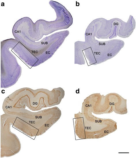
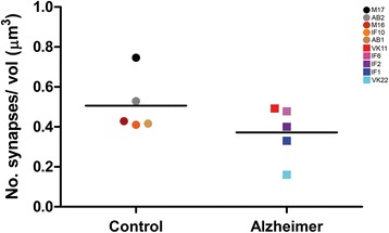
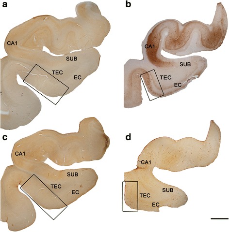
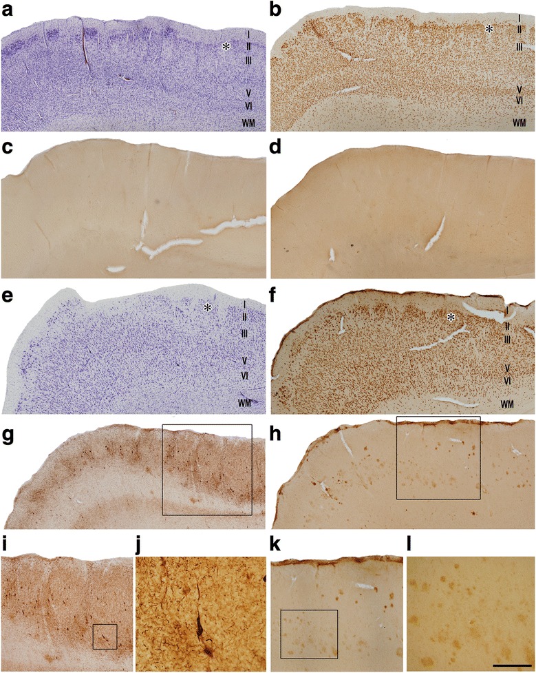
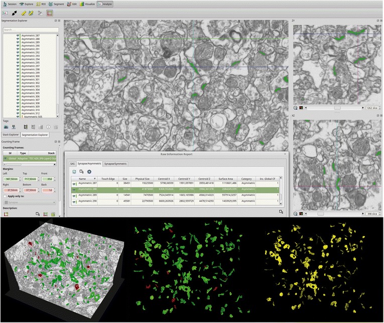
Synaptic dysfunction or loss in early stages of Alzheimer's disease (AD) is thought to be a major structural correlate of cognitive dysfunction. Early loss of episodic memory, which occurs at the early stage of AD, is closely associated with the progressive degeneration of medial temporal lobe (MTL) structures of which the transentorhinal cortex (TEC) is the first affected area. However, no ultrastructural studies have been performed in this region in human brain samples from AD patients. In the present study, we have performed a detailed three-dimensional (3D) ultrastructural analysis using focused ion beam/scanning electron microscopy (FIB/SEM) to investigate possible synaptic alterations in the TEC of patients with AD. Surprisingly, the analysis of the density, morphological features and spatial distribution of synapses in the neuropil showed no significant differences between AD and control samples. However, light microscopy studies showed that cortical thickness of the TEC was severely reduced in AD samples, but there were no changes in the volume occupied by neuronal and glial cell bodies, blood vessels, and neuropil. Thus, the present results indicate that there is a dramatic loss of absolute number of synapses, while the morphology of synaptic junctions and synaptic spatial distribution are maintained. How these changes affect cognitive impairment in AD remains to be elucidated.
在阿尔茨海默病(AD)的早期阶段,突触功能障碍或丧失被认为是认知功能障碍的主要结构相关性。在 AD 的早期阶段发生的情景记忆丧失,与内侧颞叶(MTL)结构的进行性退化密切相关,其中,外侧隔核(TEC)是第一个受影响的区域。然而,在 AD 患者的人脑样本中,尚未在该区域进行超微结构研究。在本研究中,我们使用聚焦离子束/扫描电子显微镜(FIB/SEM)进行了详细的三维(3D)超微结构分析,以研究 AD 患者 TEC 中可能存在的突触改变。令人惊讶的是,对神经突中突触密度、形态特征和空间分布的分析显示,AD 和对照组样本之间没有显著差异。然而,光学显微镜研究表明,AD 样本中 TEC 的皮质厚度严重减少,但神经元和神经胶质细胞体、血管和神经突的体积没有变化。因此,目前的结果表明存在突触绝对数量的急剧丧失,而突触连接的形态和突触的空间分布得以维持。这些变化如何影响 AD 中的认知障碍还有待阐明。