Athinoula A. Martinos Center for Biomedical Imaging, Department of Radiology, Massachusetts General Hospital and Harvard Medical School, Charlestown, Massachusetts, USA.
Harvard-MIT Division of Health Sciences and Technology, Massachusetts Institute of Technology, Cambridge, Massachusetts, USA.
J Magn Reson Imaging. 2018 Nov;48(5):1288-1296. doi: 10.1002/jmri.26000. Epub 2018 Mar 8.
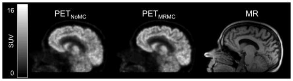
Subject motion in positron emission tomography (PET) studies leads to image blurring and artifacts; simultaneously acquired magnetic resonance imaging (MRI) data provides a means for motion correction (MC) in integrated PET/MRI scanners.
To assess the effect of realistic head motion and MR-based MC on static [ F]-fluorodeoxyglucose (FDG) PET images in dementia patients.
Observational study.
Thirty dementia subjects were recruited.
FIELD STRENGTH/SEQUENCE: 3T hybrid PET/MR scanner where EPI-based and T -weighted sequences were acquired simultaneously with the PET data.
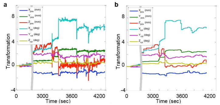
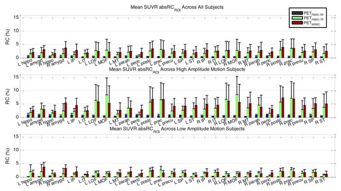
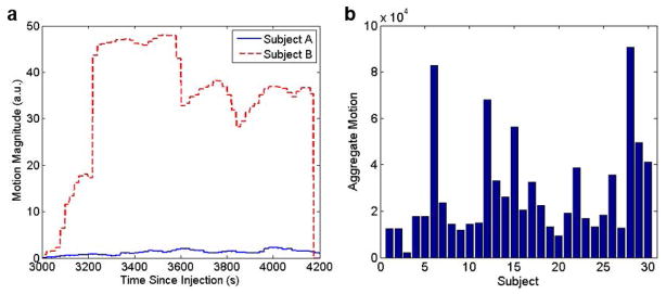
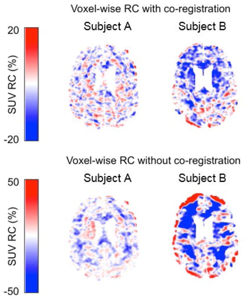
Head motion parameters estimated from high temporal resolution MR volumes were used for PET MC. The MR-based MC method was compared to PET frame-based MC methods in which motion parameters were estimated by coregistering 5-minute frames before and after accounting for the attenuation-emission mismatch. The relative changes in standardized uptake value ratios (SUVRs) between the PET volumes processed with the various MC methods, without MC, and the PET volumes with simulated motion were compared in relevant brain regions.
The absolute value of the regional SUVR relative change was assessed with pairwise paired t-tests testing at the P = 0.05 level, comparing the values obtained through different MR-based MC processing methods as well as across different motion groups. The intraregion voxelwise variability of regional SUVRs obtained through different MR-based MC processing methods was also assessed with pairwise paired t-tests testing at the P = 0.05 level.
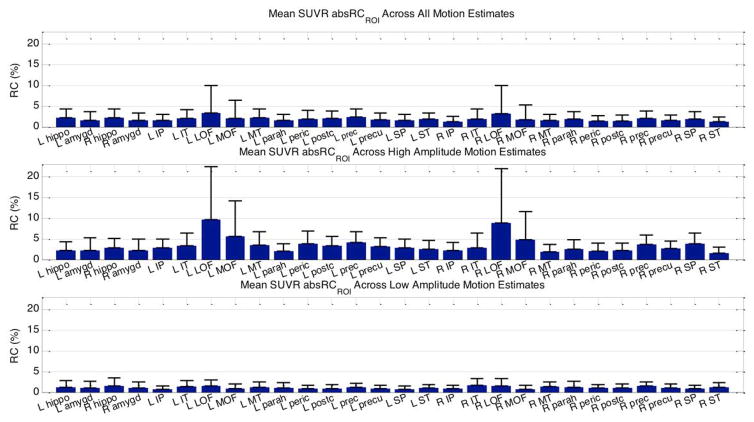
MC had a greater impact on PET data quantification in subjects with larger amplitude motion (higher than 18% in the medial orbitofrontal cortex) and greater changes were generally observed for the MR-based MC method compared to the frame-based methods. Furthermore, a mean relative change of ∼4% was observed after MC even at the group level, suggesting the importance of routinely applying this correction. The intraregion voxelwise variability of regional SUVRs was also decreased using MR-based MC. All comparisons were significant at the P = 0.05 level.
Incorporating temporally correlated MR data to account for intraframe motion has a positive impact on the FDG PET image quality and data quantification in dementia patients.
3 Technical Efficacy: Stage 1 J. Magn. Reson. Imaging 2018;47:1288-1296.
正电子发射断层扫描(PET)研究中的受试者运动导致图像模糊和伪影;同时采集的磁共振成像(MRI)数据为集成 PET/MRI 扫描仪中的运动校正(MC)提供了一种手段。
评估现实头部运动和基于 MRI 的 MC 对痴呆患者静态[F]-氟脱氧葡萄糖(FDG)PET 图像的影响。
观察性研究。
招募了 30 名痴呆患者。
场强/序列:3T 混合 PET/MR 扫描仪,其中 EPI 序列和 T1 加权序列与 PET 数据同时采集。
从高时间分辨率 MR 容积中估计的头部运动参数用于 PET MC。将基于 MRI 的 MC 方法与 PET 帧内 MC 方法进行比较,在帧内 MC 方法中,通过在考虑衰减-发射不匹配的情况下对前后 5 分钟的帧进行配准来估计运动参数。在相关脑区,比较了各种 MC 方法处理的 PET 容积、未经 MC 处理的 PET 容积和模拟运动的 PET 容积之间标准化摄取值比(SUVR)的相对变化。
使用配对 t 检验评估区域 SUVR 相对变化的绝对值,在 P=0.05 水平上进行检验,比较不同基于 MRI 的 MC 处理方法以及不同运动组获得的值。还使用配对 t 检验评估不同基于 MRI 的 MC 处理方法获得的区域 SUVR 的区域内体素变异性,在 P=0.05 水平上进行检验。
MC 对振幅运动较大(眶内侧额皮质高于 18%)的受试者的 PET 数据定量有更大的影响,与帧内 MC 方法相比,一般观察到基于 MRI 的 MC 方法的变化更大。此外,即使在组水平上,即使在 MC 后仍观察到约 4%的平均相对变化,这表明常规应用这种校正的重要性。使用基于 MRI 的 MC 还降低了区域 SUVR 的区域内体素变异性。所有比较均在 P=0.05 水平上具有统计学意义。
将时间相关的 MR 数据纳入考虑,以补偿帧内运动,对痴呆患者的 FDG PET 图像质量和数据定量有积极影响。
3 级技术功效:第 1 阶段 J. Magn. Reson. Imaging 2018;47:1288-1296.