Translational Neurology group, Department of Clinical Science, Wallenberg Neuroscience Center, Lund University, Lund, Sweden.
Department of Neurology, Scania University Hospital, Lund, Sweden.
PLoS One. 2018 Mar 8;13(3):e0194146. doi: 10.1371/journal.pone.0194146. eCollection 2018.
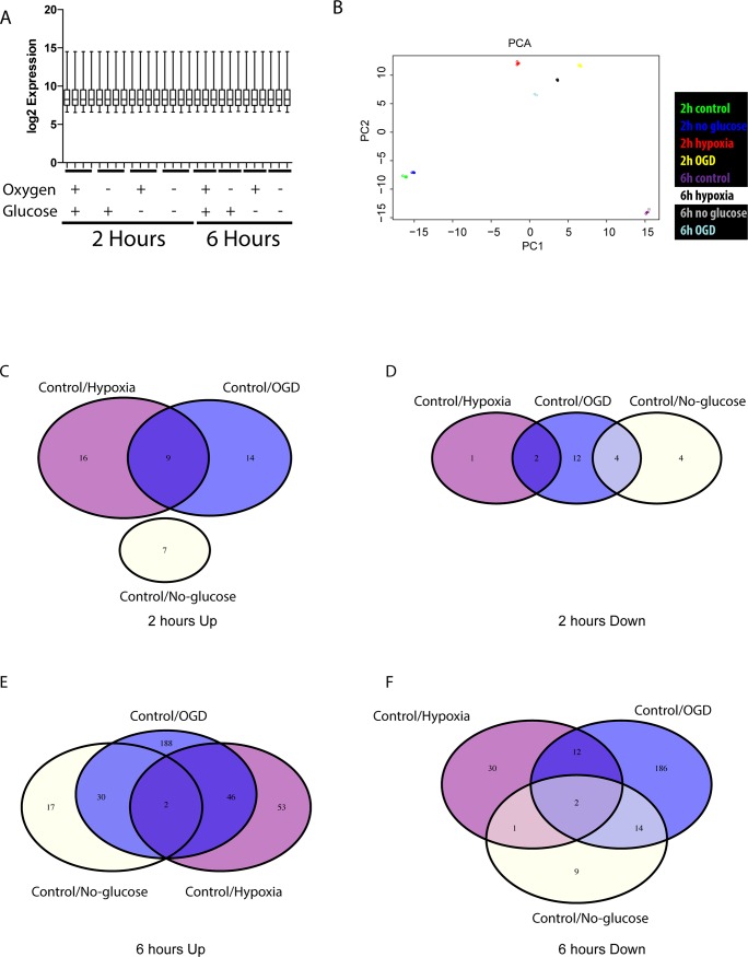
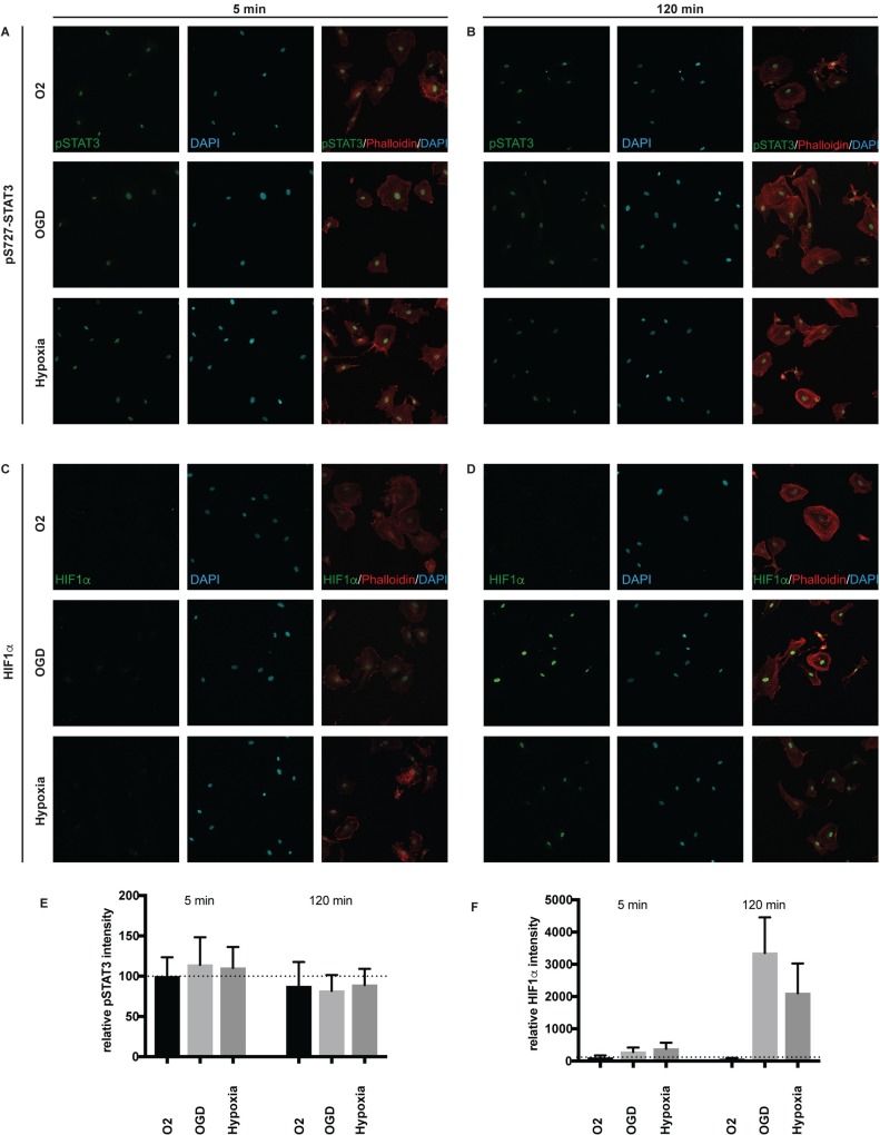
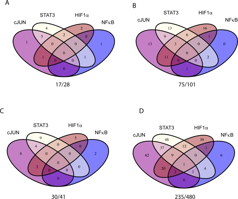
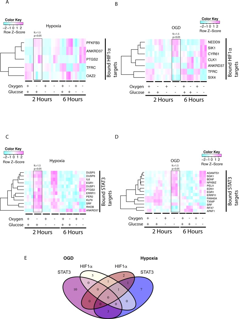
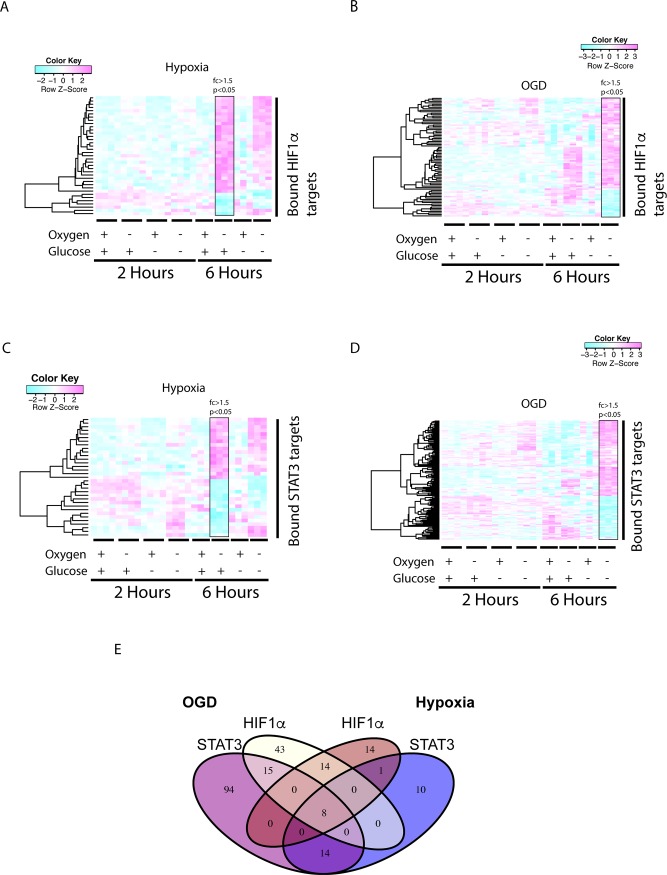
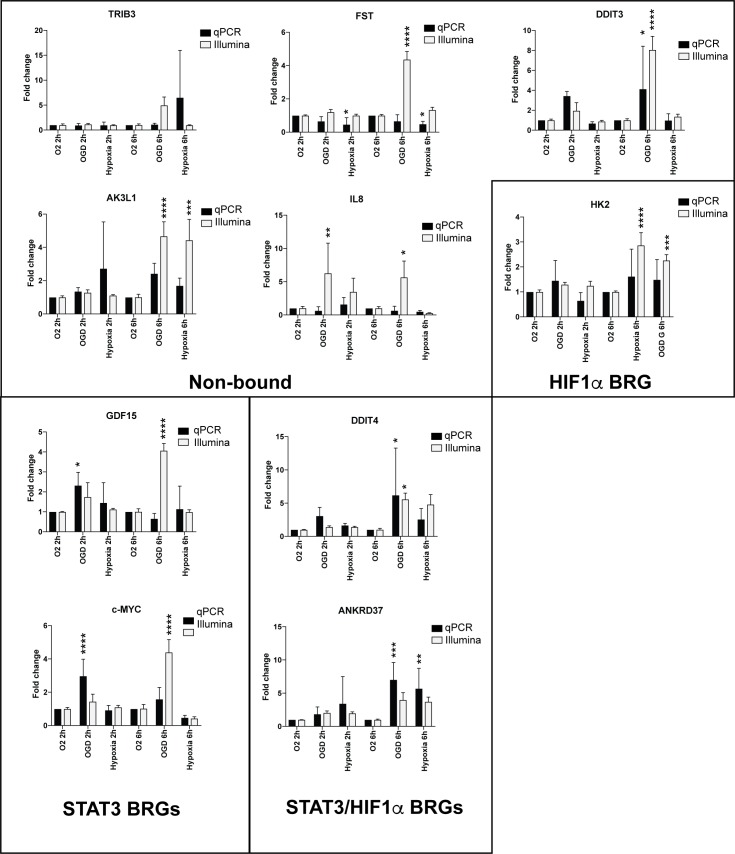
Brain pericytes are important to maintain vascular integrity of the neurovascular unit under both physiological and ischemic conditions. Ischemic stroke is known to induce an inflammatory and hypoxic response due to the lack of oxygen and glucose in the brain tissue. How this early response to ischemia is molecularly regulated in pericytes is largely unknown and may be of importance for future therapeutic targets. Here we evaluate the transcriptional responses in in vitro cultured human brain pericytes after oxygen and/or glucose deprivation. Hypoxia has been widely known to stabilise the transcription factor hypoxia inducible factor 1-alpha (HIF1α) and mediate the induction of hypoxic transcriptional programs after ischemia. However, we find that the transcription factors Jun Proto-Oncogene (c-JUN), Nuclear Factor Of Kappa Light Polypeptide Gene Enhancer In B-Cells (NFκB) and signal transducer and activator of transcription 3 (STAT3) bind genes regulated after 2hours (hs) of omitted glucose and oxygen before HIF1α. Potent HIF1α responses require 6hs of hypoxia to substantiate transcriptional regulation comparable to either c-JUN or STAT3. Phosphorylated STAT3 protein is at its highest after 5 min of oxygen and glucose (OGD) deprivation, whereas maximum HIF1α stabilisation requires 120 min. We show that STAT3 regulates angiogenic and metabolic pathways before HIF1α, suggesting that HIF1α is not the initiating trans-acting factor in the response of pericytes to ischemia.
脑周细胞对于维持神经血管单元在生理和缺血条件下的血管完整性非常重要。众所周知,缺血性中风会因脑组织缺氧和葡萄糖缺乏而引发炎症和缺氧反应。周细胞对缺血的早期反应如何在分子水平上受到调节,目前还知之甚少,这可能对未来的治疗靶点很重要。在这里,我们评估了体外培养的人脑周细胞在缺氧和/或葡萄糖剥夺后的转录反应。众所周知,缺氧会稳定转录因子缺氧诱导因子 1-α(HIF1α),并介导缺血后缺氧转录程序的诱导。然而,我们发现,转录因子 Jun 原癌基因(c-JUN)、核因子κ轻链增强子 B 细胞(NFκB)和信号转导和转录激活因子 3(STAT3)在 2 小时(hs)的葡萄糖和氧气缺失后与受调节的基因结合。强有力的 HIF1α 反应需要 6 小时的缺氧才能证明与 c-JUN 或 STAT3 相当的转录调节。磷酸化的 STAT3 蛋白在缺氧和葡萄糖剥夺(OGD)后 5 分钟达到最高水平,而最大的 HIF1α 稳定化需要 120 分钟。我们表明,STAT3 在 HIF1α 之前调节血管生成和代谢途径,这表明 HIF1α 不是周细胞对缺血反应的起始转导因子。