Molecular Biology and Biochemistry, Gottfried Schatz Research Center, Medical University of Graz, Neue Stiftingtalstraße 6/6, 8010, Graz, Austria.
BioTechMed, Graz, Austria.
Pflugers Arch. 2018 Aug;470(8):1193-1203. doi: 10.1007/s00424-018-2133-0. Epub 2018 Mar 12.
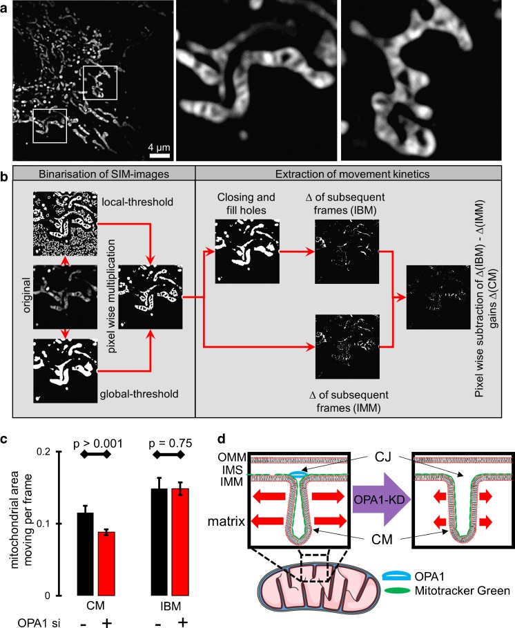
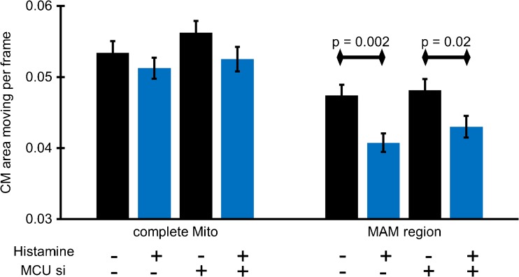
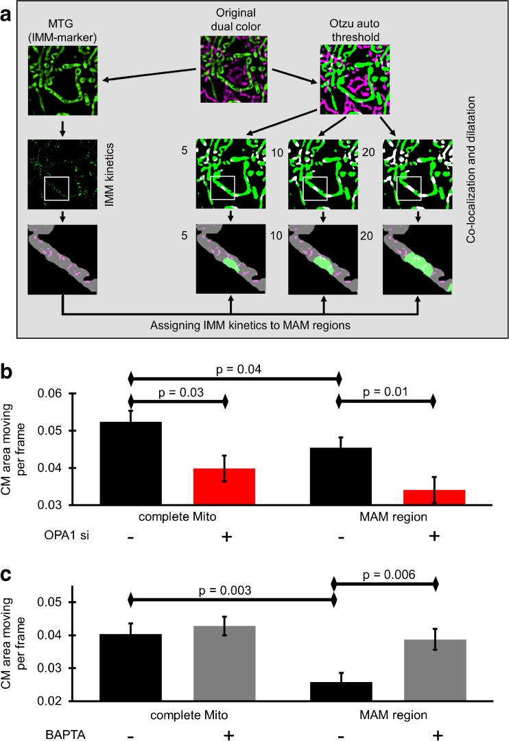
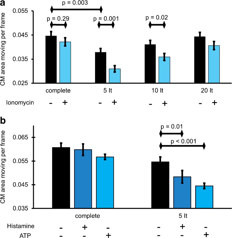
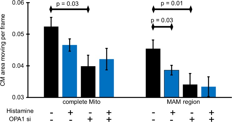
Mitochondria are multifunctional organelles that essentially contribute to cell signaling by sophisticated mechanisms of communications. Live cell imaging studies showed that mitochondria are dynamic and complex structures that form ramified networks by directed movements, fission, and fusion events. There is emerging evidence that the morphology of mitochondria determines cellular functions and vice versa. Several intracellular signaling pathways and messengers including Ca dynamically influence the architecture of mitochondria. Because electron microscopy cannot be utilized for an assessment of dynamics of mitochondrial morphology in intact cells, most studies were performed using wide-field or laser confocal fluorescence microscopies that, due to limitations of their spatial resolution, do not allow investigating sub-mitochondrial structures. Accordingly, our understanding of the dynamics of substructures of mitochondria is quite limited. Here, we present a robust super-resolution method to quantify the dynamics of mitochondrial cristae, the main substructures of the inner mitochondrial membrane, exploiting structured illumination microscopy (SIM). We observed that knockdown of the dynamin-like 120-kDa protein, which is encoded by the OPA1 gene, specifically reduces the dynamics of the mitochondrial cristae membranes (CM), while the inner boundary membrane (IBM) remained flexible. We further used dual color SIM to quantify the dynamics of CM in the junction between mitochondria and the endoplasmic reticulum (ER; mitochondrial associated membranes, MAMs). Intracellular Ca release spatially reduced CM-dynamics in MAMs. Moreover, CM-dynamics was independent from matrix Ca signal. Our data suggest that local Ca signals specifically control CM-dynamics and structure to facilitate a well-balanced functional (Ca) interplay between mitochondria and the ER.
线粒体是多功能细胞器,通过复杂的通讯机制对细胞信号转导具有重要作用。活细胞成像研究表明,线粒体是动态的、复杂的结构,通过定向运动、分裂和融合事件形成分支网络。越来越多的证据表明,线粒体的形态决定了细胞的功能,反之亦然。包括 Ca2+在内的几种细胞内信号通路和信使动态影响线粒体的结构。由于电子显微镜不能用于评估完整细胞中线粒体形态的动力学,因此大多数研究使用宽场或激光共聚焦荧光显微镜进行,由于其空间分辨率的限制,这些显微镜无法研究亚线粒体结构。因此,我们对线粒体亚结构动力学的理解相当有限。在这里,我们提出了一种稳健的超分辨率方法,利用结构照明显微镜(SIM)来定量测量线粒体嵴的动力学,线粒体嵴是线粒体内膜的主要亚结构。我们观察到,动力蛋白样 120kDa 蛋白(由 OPA1 基因编码)的敲低特异性地降低了线粒体嵴膜(CM)的动力学,而内膜边界(IBM)保持灵活。我们进一步使用双色 SIM 来定量测量线粒体与内质网(ER;线粒体相关膜,MAMs)之间连接点处的 CM 动力学。细胞内 Ca2+释放空间上减少了 MAMs 中的 CM 动力学。此外,CM 动力学与基质 Ca 信号无关。我们的数据表明,局部 Ca 信号特异性地控制 CM 动力学和结构,以促进线粒体和 ER 之间平衡的功能(Ca)相互作用。