Harvey Cushing Neuro-Oncology Laboratories, Department of Neurosurgery, Brigham and Women's Hospital, Harvard Medical School, Boston, MA 02115, USA.
Department of Neurosurgery, University Medical Center Hamburg-Eppendorf, Hamburg, Germany.
Sci Adv. 2018 Mar 7;4(3):eaar2766. doi: 10.1126/sciadv.aar2766. eCollection 2018 Mar.
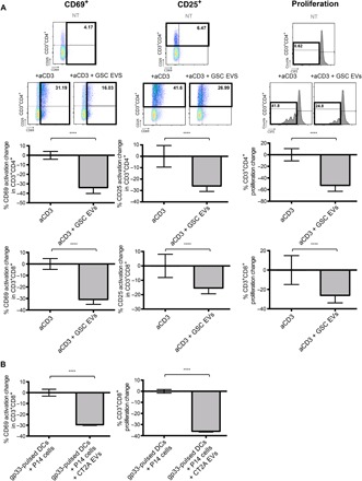
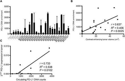
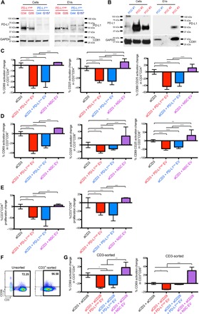
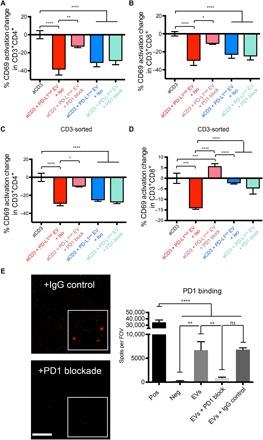
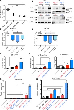
Binding of programmed death ligand-1 (PD-L1) to programmed cell death protein-1 (PD1) leads to cancer immune evasion via inhibition of T cell function. One of the defining characteristics of glioblastoma, a universally fatal brain cancer, is its profound local and systemic immunosuppression. Glioblastoma has also been shown to generate extracellular vesicles (EVs), which may play an important role in tumor progression. We thus hypothesized that glioblastoma EVs may be important mediators of immunosuppression and that PD-L1 could play a role. We show that glioblastoma EVs block T cell activation and proliferation in response to T cell receptor stimulation. PD-L1 was expressed on the surface of some, but not of all, glioblastoma-derived EVs, with the potential to directly bind to PD1. An anti-PD1 receptor blocking antibody significantly reversed the EV-mediated blockade of T cell activation but only when PD-L1 was present on EVs. When glioblastoma PD-L1 was up-regulated by IFN-γ, EVs also showed some PD-L1-dependent inhibition of T cell activation. PD-L1 expression correlated with the mesenchymal transcriptome profile and was anatomically localized in the perinecrotic and pseudopalisading niche of human glioblastoma specimens. PD-L1 DNA was present in circulating EVs from glioblastoma patients where it correlated with tumor volumes of up to 60 cm. These results suggest that PD-L1 on EVs may be another mechanism for glioblastoma to suppress antitumor immunity and support the potential of EVs as biomarkers in tumor patients.
程序性死亡配体 1(PD-L1)与程序性死亡蛋白 1(PD1)的结合导致癌症免疫逃逸,通过抑制 T 细胞功能。胶质母细胞瘤是一种普遍致命的脑癌,其定义特征之一是其深刻的局部和全身免疫抑制。胶质母细胞瘤也被证明会产生细胞外囊泡(EVs),这可能在肿瘤进展中发挥重要作用。因此,我们假设胶质母细胞瘤 EVs 可能是免疫抑制的重要介质,并且 PD-L1 可能发挥作用。我们表明,胶质母细胞瘤 EVs 可阻断 T 细胞在受到 T 细胞受体刺激时的激活和增殖。PD-L1 在一些,但不是所有的,源自胶质母细胞瘤的 EVs 表面表达,具有与 PD1 直接结合的潜力。抗 PD1 受体阻断抗体可显著逆转 EV 介导的 T 细胞激活阻断,但仅在 EV 上存在 PD-L1 时才如此。当 IFN-γ上调胶质母细胞瘤 PD-L1 时,EVs 也显示出一些依赖 PD-L1 的 T 细胞激活抑制。PD-L1 表达与间充质转录组谱相关,并在人类胶质母细胞瘤标本的坏死周围和假栅格龛中具有解剖定位。胶质母细胞瘤患者的循环 EV 中存在 PD-L1 DNA,其与肿瘤体积高达 60 cm 相关。这些结果表明,EV 上的 PD-L1 可能是胶质母细胞瘤抑制抗肿瘤免疫的另一种机制,并支持 EV 作为肿瘤患者生物标志物的潜力。