Department of Laboratory Diagnostics, The First Affiliated Hospital of USTC, Division of Life Sciences and Medicine, University of Science and Technology of China, Hefei, China.
Department of Cancer Epigenetics Program, The First Affiliated Hospital of USTC, Division of Life Sciences and Medicine, University of Science and Technology of China, Hefei, Anhui, China.
Clin Exp Immunol. 2022 May 12;207(3):307-317. doi: 10.1093/cei/uxac006.
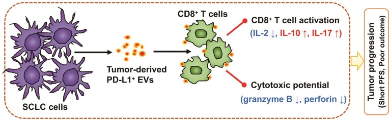
Programmed death ligand-1 (PD-L1) is expressed on the surface of tumor cells and binds to programmed cell death protein-1 (PD1) on the surface of T cells, leading to cancer immune evasion via inhibition of T-cell function. One of the characteristics of small cell lung cancer (SCLC) is its ineffective anti-tumor immune response and highly immunosuppressive status in the tumor microenvironment. SCLC cells have been shown to generate extracellular vesicles (EVs), which may play an important role in tumor progression. We thus hypothesized that SCLC EVs may be important mediators of immunosuppression and that PD-L1 could play a role. Herein, we showed that PD-L1 was expressed on the surface of SCLC-derived EVs, with the potential to directly bind to PD1. Experimentally, we further showed that EVs secreted by SCLC cells can inhibit CD8+ T-cell activation and cytokine production in vitro in response to T-cell receptor stimulation. Importantly, an anti-PD-L1 blocking antibody significantly reversed the EV-mediated inhibition of CD8+ T-cell activation. Furthermore, we performed a retrospective study of patients with SCLC to determine the prognostic value of PD-L1 harvested from plasm circulating EVs. The results showed that EVs containing PD-L1 was an independent prognostic factor and significantly correlated with progression-free survival. Together, these results indicate that EVs containing PD-L1 can be served as a diagnostic biomarker for predicting the effectiveness of therapy, as well as a new strategy to enhance T-cell-mediated immunotherapy against SCLC cancers.
程序性死亡配体-1(PD-L1)表达于肿瘤细胞表面,与 T 细胞表面的程序性死亡蛋白-1(PD1)结合,通过抑制 T 细胞功能导致癌症免疫逃逸。小细胞肺癌(SCLC)的特征之一是其抗肿瘤免疫反应无效,肿瘤微环境中具有高度免疫抑制状态。已经表明 SCLC 细胞会产生细胞外囊泡(EVs),这可能在肿瘤进展中发挥重要作用。因此,我们假设 SCLC EVs 可能是免疫抑制的重要介质,并且 PD-L1 可能发挥作用。在此,我们表明 PD-L1 表达于 SCLC 衍生的 EVs 表面,具有直接与 PD1 结合的潜力。实验上,我们进一步表明 SCLC 细胞分泌的 EVs 可以抑制体外 T 细胞受体刺激后 CD8+T 细胞的激活和细胞因子产生。重要的是,抗 PD-L1 阻断抗体显著逆转了 EV 介导的 CD8+T 细胞激活抑制。此外,我们对 SCLC 患者进行了回顾性研究,以确定从血浆循环 EVs 中提取的 PD-L1 的预后价值。结果表明,含有 PD-L1 的 EVs 是一个独立的预后因素,与无进展生存期显著相关。综上所述,这些结果表明,含有 PD-L1 的 EVs 可以作为预测治疗效果的诊断生物标志物,也是增强针对 SCLC 癌症的 T 细胞介导免疫疗法的新策略。